Download
1 / 58
580 likes | 700 Views
Blood. Ch 17. Blood. Artery. White blood cells. Platelets. Red blood cells. Function Blood. Deliver O2 Remove metabolic wastes Maintain temperature, pH, and fluid volume Protection from blood loss- platelets Prevent infection- antibodies and WBC Transport hormones. Blood.

E N D
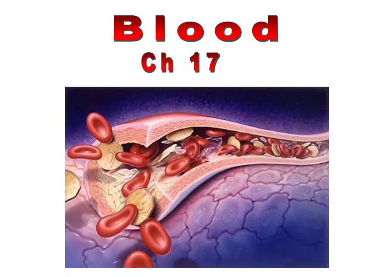
Blood Ch 17
Blood Artery White blood cells Platelets Red blood cells
Function Blood • Deliver O2 • Remove metabolic wastes • Maintain temperature, pH, and fluid volume • Protection from blood loss- platelets • Prevent infection- antibodies and WBC • Transport hormones
Blood Plasma-55% Buffy coat-<1% Formed elements-45%
Blood Plasma Components-55% 90% Water 8% Solutes: • Proteins Albumin (60 %) Alpha and Beta Globulins Gamma Globulins fibrinogens • Gas • Electrolytes
Blood Plasma Components • Organic Nutrients Carbohydrates Amino Acids Lipids Vitamins • Hormones • Metabolic waste CO2 Urea
Buffy Coat- <1% • Leukocytes • Platelets
Formed Elements of the Blood-45% • Erythrocytes (red blood cells) • Leukocytes (white blood cells) • Platelets (thrombocytes)
Erythrocyte7.5m in dia ·Anucleate- so can't reproduce; however, repro in red bone marrow ·Hematopoiesis- production of RBC ·Function- transport respiratory gases ·Hemoglobin- quaternary structure, 2 chains and 2 chains ·Lack mitochondria. Why? ·1 RBC contains 280 million hemoglobin molecules ·Men- 5 million cells/mm3 ·Women- 4.5 million cells/mm3 ·Life span 100-120 days and then destroyed in spleen (RBC graveyard)
Hematopoiesis • Hematopoiesis (hemopoiesis): blood cell formation • Occurs in red bone marrow of axial skeleton, girdles and proximal epiphyses of humerus and femur
Hematopoiesis • Hemocytoblasts (hematopoietic stem cells) • Give rise to all formed elements • Hormones and growth factors push the cell toward a specific pathway of blood cell development • New blood cells enter blood sinusoids
Erythropoiesis • Erythropoiesis: red blood cell production • A hemocytoblast is transformed into a proerythroblast • Proerythroblasts develop into early erythroblasts
Erythropoiesis • Phases in development • Ribosome synthesis • Hemoglobin accumulation • Ejection of the nucleus and formation of reticulocytes • Reticulocytes then become mature erythrocytes
Stem cell Committed cell Developmental pathway Phase 1 Ribosome synthesis Phase 2 Hemoglobin accumulation Phase 3 Ejection of nucleus Reticulo- cyte Erythro- cyte Proerythro- blast Early erythroblast Late erythroblast Normoblast Hemocytoblast Figure 17.5
Regulation of Erythropoiesis • Too few RBCs leads to tissue hypoxia • Too many RBCs increases blood viscosity • Balance between RBC production and destruction depends on • Hormonal controls • Adequate supplies of iron, amino acids, and B vitamins
Hormonal Control of Erythropoiesis • Erythropoietin (EPO) • Direct stimulus for erythropoiesis • Released by the kidneys in response to hypoxia
Hormonal Control of Erythropoiesis • Causes of hypoxia • Hemorrhage or increased RBC destruction reduces RBC numbers • Insufficient hemoglobin (e.g., iron deficiency) • Reduced availability of O2 (e.g., high altitudes)
Hormonal Control of Erythropoiesis • Effects of EPO • More rapid maturation of committed bone marrow cells • Increased circulating reticulocyte count in 1–2 days • Testosterone also enhances EPO production, resulting in higher RBC counts in males
RBC Diseases Anemia- when blood has low O2 carrying capacity; insufficient RBC or iron deficiency. Factors that can cause anemia- exercise, B12 deficiency
RBC Diseases Sickle-cell anemia- • HbS results from a change in just one of the 287 amino acids in the chain in the globin molecule. • Found in 1 out of 400 African Americans. • Homozygous for sickle-cell is deadly, but in malaria infested countries, the heterozygous condition is beneficial.
Genetics of Sickle Cell Anemia Genetics of Sickle Cell Anemia
RBC Diseases Polycythemia- excess of erythrocytes, viscosity of blood; 8-11 million cells/mm3 Usually caused by cancer; however, naturally occurs at high elevations Blood doping- in athletesremove blood 2 days before event and then replace it- banned by Olympics.
Types of Leukocytes 4,000-11,000 cells/mm 3 Never let monkeys eat bananas Granulocytes Neutrophils- 40-70% Eosinophils- 1-4% Basophils- <1% Agranulocytes Monocytes- 4-8% Lymphocytes- 20-45%
Lymphocyte Eosinophil Basophil platelet Neutrophil Monocyte
Diapedesis Leukocyte Squeezing Through Capillary Wall
WBC Diseases • Leukopenia • Abnormally low WBC count—drug induced • Leukemias • Cancerous conditions involving WBCs • Named according to the abnormal WBC clone involved • Mononucleosis • highly contagious viral disease caused by Epstein-Barr virus; excessive # of agranulocytes; fatigue, sore throat, recover in a few weeks
Platelets • Small fragments of megakaryocytes • Formation is regulated by thrombopoietin • Blue-staining outer region, purple granules • Granules contain serotonin, Ca2+, enzymes, ADP, and platelet-derived growth factor (PDGF)
Stem cell Developmental pathway Hemocyto- blast Promegakaryocyte Megakaryoblast Megakaryocyte Platelets Figure 17.12
Platelet Plug Clotting Factors Hemostasis- stoppage of bleeding Platelets: 250,000-500,000 cells/mm3 Tissue Damage
Hemostasis: • Vessel injury 2. Vascular spasm 3. Platelet plug formation 4. Coagulation
Clotting Factors thromboplastin Prothrombin Thrombin Fibrinogen Fibrin Hemostasis(+ feedback) Traps RBC & platelets Platelets release thromboplastin
Blood Clot RBC Platelet Fibrin thread
Disorders of Hemostasis • Thromboembolytic disorders: undesirable clot formation • Bleeding disorders: abnormalities that prevent normal clot formation
Thromboembolytic Conditions • Thrombus: clot that develops and persists in an unbroken blood vessel • May block circulation, leading to tissue death • Embolus: a thrombus freely floating in the blood stream • Pulmonary emboli impair the ability of the body to obtain oxygen • Cerebral emboli can cause strokes
Thromboembolytic Conditions • Prevented by • Aspirin • Antiprostaglandin that inhibits thromboxane A2 • Heparin • Anticoagulant used clinically for pre- and postoperative cardiac care • Warfarin • Used for those prone to atrial fibrillation
Bleeding Disorders • Thrombocytosis- too many platelets due to inflammation, infection or cancer • Thrombocytopenia- too few platelets • causes spontaneous bleeding • due to suppression or destruction of bone marrow (e.g., malignancy, radiation) • Platelet count <50,000/mm3 is diagnostic • Treated with transfusion of concentrated platelets
Bleeding Disorders • Impaired liver function • Inability to synthesize procoagulants • Causes include vitamin K deficiency, hepatitis, and cirrhosis • Liver disease can also prevent the liver from producing bile, impairing fat and vitamin K absorption
Bleeding Disorders • Hemophilias include several similar hereditary bleeding disorders • Symptoms include prolonged bleeding, especially into joint cavities • Treated with plasma transfusions and injection of missing factors
Hemophiliac- a sex-linked recessive trait, primarily carried by males (x chromosome)
Blood Types Type A Type B Type AB Type O
Blood Typing Blood type is based on the presence of 2 major antigens in RBC membranes-- A and B Blood type Antigen Antibody A A anti-B B B anti-A A & B AB no anti body Neither A or B O anti-A and anti-B Antigen- protein on the surface of a RBC membrane Antibody- proteins made by lymphocytes in plasma which are made in response to the presence of antigens. They attack foreign antigens, which result in clumping (agglutination)
b b b b b b b Type A
a a a a a a a Type B
a a a b a a a b b b Type O