Download
1 / 5
50 likes | 384 Views
The Spinal Cord. Lecture 3. Location. The spinal cord is contained in and protected by the vertebrae. In the embryo, the spinal cord occupies the entire spinal canal, extending down into the coccyx portion of the vertebral column.

E N D
The Spinal Cord Lecture 3
Location • The spinal cord is contained in and protected by the vertebrae. • In the embryo, the spinal cord occupies the entire spinal canal, extending down into the coccyx portion of the vertebral column. • The column of bone grows much more rapidly than the nerve tissue of the cord, so that in adults, the cord ends in the region just below the area to which the last rib attaches (between the 1st and 2nd lumbar vertebrae).
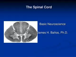
Structure • The spinal cord has a small, irregularly shaped internal section that consists of gray matter (nerve cell bodies) and a larger area surrounding this gray matter that consists of white matter (nerve cell fibers). • The gray matter is arranged in two pairs of columns, one dorsally and one ventrally, to form a butterfly shaped region in the middle of the spinal cord. The two pairs of columns are called the dorsal and ventral horns. • In the center of the gray matter is a small channel, the central canal, which contains cerebrospinal fluid. • The white matter consists of thousands of myelinated axons arranged in three areas external to the gray matter on each side.
Functions • Linking the Spinal Nerves to the Brain • The white matter of the spinal cord is divided into tracts that convey impulses to and from the brain. • Sensory impulses from the peripheral receptors enter the dorsal horn of the spinal cord. • They are then transmitted upward toward the brain in ascending tracts of white matter. • Motor impulses traveling down from the brain are carried in descending tracts until they exit through the ventral horn of the gray matter to reach an effector.
Reflex Activities • A reflex is a rapid, simple, and automatic response involving very few neurons. • Reflexes are specific; a given stimulus always produces the same response. • A simple reflex arc that passes through the spinal cord alone and does not involve the brain is termed a spinal reflex. • One example of a spinal reflex is the stretch reflex. • This is when a muscle is stretched and responds by contracting. • If you tap the patellar tendon, the muscle of the quadriceps femoris contracts, eliciting a knee-jerk reflex. • Such stretch reflexes may be evoked by appropriate tapping of most large muscle. Because reflexes are simple and predictable, they are used to test the condition of the Nervous System.