Download
1 / 16
170 likes | 333 Views
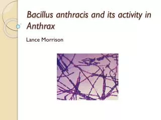
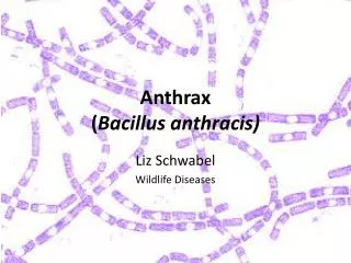
/2. Bacillus anthracis .. Cutaneous Black Lesions.. Clostridium perfingens and other sp. : Necrotizing Fasciitis.. Myonecrosis , Cellulitis , tissues putrefaction, gas production/ Gas gangrene.. Surgical/Traumatic wound.. Skin- Subcutaneous (Mixed Infection).. Specific Enzymes & Exotoxins

E N D
/2 • Bacillus anthracis..Cutaneous Black Lesions.. • Clostridium perfingensand other sp.: Necrotizing Fasciitis.. Myonecrosis, Cellulitis, tissues putrefaction, gas production/ Gas gangrene.. Surgical/Traumatic wound.. Skin- Subcutaneous (Mixed Infection).. Specific Enzymes & Exotoxins • BorreliaBurgdorferi: Lyme disease .. Transmitted by Tick/ Insect bites.. Incub. 1-3 weeks.. Annular Rash.. Chronic Skin Lesion.. Cardiac & Neurological Abnormality.. Arthritis.. Endemic USA, China, Japan • Bartonella species: G-ve bacilli BartonellosisCat Scratch Fever..followed Cat scratch or bite..Skin lesions.. Subacute regional lymphadenitis..Septicemia.
Tuberculosis-Leprosy-1 • Cutaneous Tuberculosis(TB).. Cutaneous TB is a relatively uncommon form of extra-pulmonary TB.. • Rare M. tuberculosis.. Common M. marinum-ulcerans.. Low Temperature..Water.. Skin Lesions.. Chronic cutaneous ulcer.. Small granulomas Follow skin injury..Trauma. • Leprosy:Chronic bacterial infectioncaused by M. leprae.. Itprimarily affects cold body sites skin, mucous membranes.. peripheral nerves ..nose, ears, eye lids and testes. • characterized by multiple skin lesions accompanied first by sensation loss/ anesthesia.. sensory loss in the affected areas, toes, finger tips, tissuedestructions.
Leprosy-3 3/ • Lebrosy can affect people of all races around the world. However, it is most common in warm, wet areas in the tropics and subtropics. • In most cases, it is spread through long-term contact with a person who has the disease but has not been treated. • Most people will never develop the disease even if they are exposed to the bacteria.. have a natural immunity to leprosy. • Worldwide prevalence is reported to be around 5.5 million, with 80% of these cases found in 5 countries: India, Indonesia, Myanmar, Brazil and Nigeria.
Clinical Leprosy-4 • Infection incubation period range from 6 months - 40 years or longer. usually begins in the extremities • Leprosy formsdepend on the person's immune response to the infection. • There are several forms of leprosy: • Tuberculoid form..Mild Form.. Few AF Bacilli, Lepromin skin test +ve, Presence nerve sensation • lepromatous type Severe form.. Numerous Acid-fast bacilli, Loss nerve sensation.. Lepromin skin test -ve
Diagnosis & Treatment • Lab Diagnosis: A skin biopsy may show characteristic granulomas (mixed inflammatory cell infiltrate in the deeper layers of the skin, the dermis) with involvement of the nerves. • Presence Acid fast bacilli.. number of bacilli visible depending on the type of leprosy.. No Culture.. No Protected Vaccine available.. BCG may help & reduce the severity of disease • Treatment:Dapsone, Rifampin, Clofazimine. Life-long Treatment ..No Cure but Less Tissue Damage and Spread of Infection.
Common Fungal Skin Infection-1 • Superficial & Cutaneous Mycosis: Invade only dead tissues of the skin.. keratinized body tissues.. Skin, Hair, Nails. causes skin peeling, redness, itching, burning.. less blisters and sores. • Malnourishment, poor hygiene, suppressed immunity & warm moist climate may increase the incidence fungal skin infection • Dermatophytes:Trichopyhton, Microsporum, Epidermatophyton spp., Yeast forms Piytrosporum, Trichosporons..present in hair follicles & skin folding. • Transmission: Usually from person to person or animal to person.. dust particles..common more with chronic skin disorders.
Skin Fungal Infection-2 • Tineacapitis:Hair follicles, scalp circular patches.. Scaling,Hair Loss..Children..Rare adults • Tineacorporis: Skin annular-erythematic lesions, Vesicles, Scaling.. Itching.. Rash.. All Ages.. Mostly caused by Dermatophytes ..rarely mixed with Yeast • Tineapedis:Red vesicles.. Interdigital spaces, web lesions, Toes, Plantar surface.. Feet, Itching.. Chroniclesions..Wearing tight shoes/socks, increased feet sweating.. More in Adults than children.. Cased by all Dermatophytes. • Tineacruris: Pelvic area.. Groin.. Erythematic Lesions, Itching, Chronic.. more common in male young adults..mostly Epidermophytonspp
Skin Fungal Infection-3 • Tina unguium(Onychomycosis): Mostly caused by Trichophyton ,Microsporum.. less Candida..fingernails & toenails. Nails become colorless/dark colored, thicken, disfigure and brittle..Diabetes • Psoriasisis a common skin disorder produces thick red plaques covered with silvery scales..can affect the nails, scalp, skin and joints..not caused by fungus and not transmitted to others. • Eczema develops due to multiple immunological & other medical conditions.. Skin becomes inflamed or irritated..No infectious agent involved. • Aspergillus & Cryptococcus spp. Rare cause localised skin or nail..
Skin Fungal Infection-3 • TineaVersicolor/Pityriasis:Malasseziafurfur / Piytrosporumfolliculitis.. Lipophilic Yeast ..difficult to culture in Labs. Part skin flora.. Endogenous infection.. Skin Moist-Folded Area.. Discoloration.. Red Spots.. Mostly Face-Neck FingerTrunk..Mild..rarely Chronic, Stress conditions, UV-Light, Common in young adults. • Head dundruff, Seborrheic dermatitis. • White & Black Piedra..Trichosporon spp., Soft to hard nodules. scalp hair & hair shaft , skin face , any body part.
Yeat skin infection • Candidasis: C. albicans, C. glabrata, C. tropicalis.. Other spp. Endogenous infection..moist folds of skin.. Lesions, finger nails, toenails, Finger webs.. Diabetes, immuno-compromessed.. more common in Infant & women.. Candida infections can look just like other types of dermatitis /eczema or skin allergy. itching, redness..infection • Blasmycosis: Blastomycesdermatitidis & Histoplasmosis : Histoplasmacapsulatum.. Dimorphic Fungi.. Soil ..Spore Inhalation.. Respiratory infection.. Systemic Infection.. Complications: Skin ulcerations/lesions Granulomas..causes severe damages..common USA, Canada
Lab diagnosis-4 • Direct microscopic examinationof skin scales dissolved in a 10 % solution potassium hydroxide (KOH).. demonstrating the fungus as small Filaments / Yeast like structures. • Culture:Sabouraud Dextrose agar, Incubation at room temperature & 37 C for 2-6 Weeks. . Slow growth for Dermatophytes..Rapid growth Candida. • ChromCandida agar.. used for rapid identification of common Candida species. • Treatment:Most skin infections respond very well to topical antifungaldrugs..Less systemic drug.. interact with Ergosterol ..causing Fungal Cell membrane disruption.. Imidazole drugs..miconazole, clotrimazole, econazole, ketoconazole, fluconazole