Download
1 / 22
250 likes | 605 Views
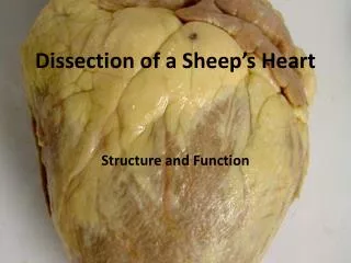
Mammal Heart Dissection. External Structure of the Heart. The Pericardium. Examine the heart and locate the thin membrane or pericardium that still covers the heart. The pericardium or pericardial sac, is a double-layered closed sac that surrounds the heart and anchors it.

E N D
The Pericardium • Examine the heart and locate the thin membrane or pericardium that still covers the heart. • The pericardium or pericardial sac, is a double-layered closed sac that surrounds the heart and anchors it. • The pericardium consists of two tissues layers - the visceral pericardium that covers the surface of the heart & the parietal pericardium covering the inner surface of the parietal sac. • The slender gap between the parietal & visceral surfaces is the pericardial cavity & is filled with fluid to reduce friction between the layers as the heart pumps.
The Myocardium • Carefully remove the pericardium. • Located below the pericardium is the muscle of your heart called the myocardium. • Most of the myocardium is located in the lower two chambers of the heart called ventricles.
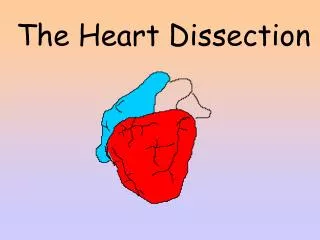
Orientation of the Heart • Locate the tip of the heart or the apex. • Only the left ventricle extends all the way to the apex. • Place the heart in the dissecting pan so that the front or ventral side is towards you ( the major blood vessels are on the top and the apex is down). • The front of the heart is recognized by a groove that extends from the right side of the broad end of the heart diagonally to a point above & to your left of the apex. • The heart is now in the pan in the position it would be in a body as you face the body.
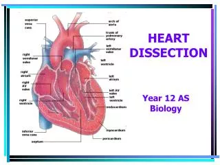
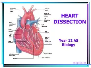
Label the Following • Apex • Left atria - upper chamber to your right • Left ventricle - lower chamber to your right • Right atria - upper chamber to your left • Right ventricle - lower chamber to your left
Major Arteries of the Heart Label the Following: • Coronary artery - this blood vessel lies in the groove on the front of the heart & it branches over the front & the back side of the heart to supply fresh blood with oxygen & nutrients to the heart muscle itself. • Pulmonary artery - this blood vessel branches & carries blood to the lungs to receive oxygen & can be found curving out of the right ventricle (upper chamber to your left) • Aorta - major vessel located near the right atria & just behind the pulmonary arteries to the lungs. Locate the curved part of this vessel known as the aortic arch. Branching from the aortic arch is a large artery that supplies blood to the upper body.
Major Veins of the Heart Label the Following: • Pulmonary Veins - these vessels return oxygenated blood from the right & left lungs to the left atrium (upper chamber on your right) • Inferior & Superior Vena Cava - these two blood vessels are located on your left of the heart and connect to the right atrium (upper chamber on your left). Deoxygenated blood enters the body through these vessels into the right receiving chamber. Use your probe to feel down into the right atrium. These vessels do not contain valves to control blood flow.
6 *2, 3 and 4 are all branches of the same artery 7
Aorta 2-4 Coronary Arteries 5. Pulmonary Artery 6. Superior Vena Cava 7. Inferior Vena Cava 6 7
Incision #1 • Use scissors to cut through the side of the pulmonary artery and continue cutting down into the wall of the right ventricle. Be careful to just cut deep enough to go through the wall of the heart chamber. (Your cutting line should be above & parallel to the groove of the coronary artery.) • With your fingers, push open the heart at the cut to examine the internal structure. If there is dried blood inside the chambers, rinse out the heart.
The Right Atrium • Locate the right atrium. Notice the thinner muscular wall of this receiving chamber. • Find where the inferior & superior vena cava enter this chamber & notice the lack of valves.
Tricuspid Valve • Locate the valve that between the right atrium and ventricle. • This valve allows blood flow from the right atrium into the right ventricle. • This is called the tricuspid valve. The valve consists of three leaflets & has long fibers of connective tissue called chordaetendinaethat attach it to papillary muscles of the heart. • When the heart begins to contract (systole phase), ventricular pressure increases until it is greater than the pressure in the atrium causing the tricuspid to snap closed.
The Right Ventricle • Use your fingers to feel the thickness of the right ventricle and its smooth lining. Also note the network of irregular muscular cords on the inner wall of this chamber. • Find the septum on the right side of the right ventricle. This thick muscular wall separates the right & left pumping ventricles from each other. • Inside the right ventricle, locate the pulmonary artery that carries blood away from this chamber.
Pulmonary Semilunar Valve • Find the one-way valve that controls blood flow away from the right ventricle at the entrance to this blood vessel.
Left Atrium • Examine the left atrium. • Find the openings of the pulmonary veins from the lungs
Valves of the Left Heart • Observe the one-way, semi-lunar valves at the entrance to the pulmonary veins. • Inside this chamber, look for the valve that controls blood flow between the upper left atrium and lower left ventricle. • This valve is called the bicuspid or mitral valve. This valve consists of two leaflets & blood flows from the left atrium into the left ventricle.
The Left Ventricle • Examine the left ventricle. • Notice the thickness of the ventricular wall. • This heart chamber is responsible for pumping blood throughout the body.
Aortic Semilunar Valve • Count the three flaps or leaflets on this valve leading from the left ventricle into the aorta and note their half-moon shape.