Download
1 / 199
2.11k likes | 2.69k Views
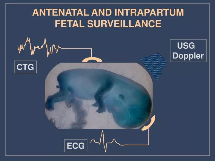
ANTENATAL AND INTRAPARTUM FETAL SURVEILLANCE. USG Doppler. C TG. E C G. MAJOR CAUSES OF PERINATAL LOSS Fetal abnormality Preterm delivery Chronic utero – placental insufficiency Acute Hypoxia Unexplained intrauterine death Others. MODALITIES OF ANTENATAL FETAL

E N D
ANTENATAL AND INTRAPARTUM FETAL SURVEILLANCE USG Doppler CTG ECG
MAJOR CAUSES OF PERINATAL LOSS Fetal abnormality Preterm delivery Chronic utero – placental insufficiency Acute Hypoxia Unexplained intrauterine death Others
MODALITIES OF ANTENATAL FETAL SURVEILLANCE Detection of fetal abnormalities Assessment of fetal growth Monitoring of fetal well-being Others
APPROACH TO ANTENATAL MONITORING OF FETAL GROWTH AND WELL-BEING Confirmation of gestational age Clinical methods of monitoring Investigations
DYNAMIC PARAMETERS To monitor fetal dynamic response to chronic utero-placental insufficiency AFI – renal perfusion Umbilical arterial flow (A/B ratio) – fetal peripheral and placental circulation Cerebral arterial flow (MCA) – head sparing effect
FETAL MONITORING Fetal distress is defined in terms of the manifestation of the fetal hypoxia (by changes in the fetal heart rate FHR or fetal blood pH) SIGNIFICANCES OF FETAL DISTRESS Hypoxic damage to the foetus is difficult to quantify, but the effect can be devastating. - neurological abnormalities - cerebral palsy and mental retardations - fetal death – results from severe intrapartum asphyxia
BASIC DEFINITIONS HYPOXAEMIA- decrease in the oxygen content of the arterial blood alone HYPOXIA- decrease in the oxygen content that affects the peripheral tissues ASPHYXIA- general oxygen deficiency that affects the high priority organs as well
PATHOPHYSIOLOGY OF FETAL HYPOXIA In the absence of stress, the fetus is neither acidotic nor hypoxic. An adequate delivery of oxygen to the tissues occurs despite the low fetal arterial partial pressure of oxygen (pO2). The transfer of oxygen across the placenta to the fetus is enhanced by following mechanisms : - fetal cardiac output and systematic blood flow rates are higher than those of the adult. - the affinity of fetal blood for the oxygen and - the fetal oxygen – caring capacity, both of which are greater than those of an adult
PATHOPHYSIOLOGY OF CHRONIC FETAL COMPROMISE utero-placental insufficiency reduced pO2 to fetal CNS redistribution of fetal cardiac output perfusion to CNS & heart perfusion to peripheral & viscera renal perfusion visceral circulation OLIGOHYDRAMNIOS bowel distension umbilical arterial resistance meconium peritonitis necrotising enterocolitis etc
Sequelae of Chronic Oligohydramnios 1. fetal demise 2. pulmonary hypoplasia 3. facial deformities 4. skeletal deformities
A decreased amniotic fluid volume is frequently one of the first clues to an underlying fetal abnormality. The sonographer/sonologist should, therefore, have a basic understanding of the mechanisms responsible for normal amniotic fluid production. Once the derivation of amniotic fluid is understood, the potential mechanisms that can result in oligohydramnios can be better appreciated.
Etiology of Oligohydramnios 1. intrauterine growth restriction 2. post-term pregnancies 3. preterm rupture of the membranes 4. fetal anomalies and/or aneuploidy 5. iatrogenic
30 week gestation. A single deepest pocket of amniotic fluid (7 cm), indicating a normal amniotic fluid volume. Visually normal amniotic fluid volume at 18 weeks' gestation. 1.8 cm pocket of amniotic fluid indicating oligohydramnios. The color box confirms that umbilical cord is not present in the pockets of amniotic Subjective assessment of amniotic fluid volume. 20 week fetus with a unilateral multicystic kidney and congenital absence of the other kidney, resulting in anhydramnios
Oligohydramnios due to bilateral renal agenesis. Transabdominal ultrasound at 20 weeks' gestation.
An amniotic fluid index of 4.2 cm, indicating oligohydramnios there is an absence of amniotic fluid in the upper quadrants. The color box to the right of the image indicates the presence of umbilical cord.
19 week fetus with Turner's syndrome, cystic hygroma (arrows) and oligohydramnios. 15 week fetus with posterior urethral valves. The bladder (b) is massively distended. Enlarged "key-hole" bladder associated with posterior urethral valves.
Amniotic fluid has a number of important roles in embryo/fetal development: 1. Permitting fetal movement and the development of the musculoskeletal system. 2. Swallowing of amniotic fluid enhances the growth and development of the gastrointestinal tract. 3. The ingestion of amniotic fluid provides some fetal nutrition and essential nutrients.
Amniotic fluid has a number of important roles in embryo/fetal development: 4. Amniotic fluid volume maintains amniotic fluid pressure thereby reducing the loss of lung liquid an essential component to pulmonary development. 5. Protects the fetus from external trauma. 6. Protects the umbilical cord from compression. 7. It's constant temperature helps to maintain the embryo's body temperature. 8. It's bacteristatic properties reduces the potential for infection.
STRESSED FETUS When perfusions is decreased because of impaired uterine or umbilical blood flow , the transfer of oxygen to the fetus is diminished, and the results is an accumulations of carbon dioxide in the fetus. 1. Increased carbon dioxide causes an increase in the partial pressure of carbon dioxide (pCO2) and a concomitant fall in pH, analogous to adult respiratory acidosis. 2. Continued hypoxia deprives the fetus of sufficient oxygen to perform the aerobic reactions, resulting in buildup of organic acids with the accumulation of pyruvic and lactic acids resulting in metabolic acidosis
STRESSED FETUS 3. Transient decreases in the fetal or uterine perfusions usually cause a short-lived respiratory acidosis, whereas more prolonged or profound decreases results in a combined respiratory and metabolic acidosis 4. fetal oxygen deprivation usually results in the FHR or fetal bradycardia
FETAL DEFENCE MECHANISMS • Increased tissue oxygen extraction • Reduced non-essential activity • Increased sympathetic activity • Redistribution of blood flow • Anaerobic metabolism with the metabolism • of blood sugar – glucolysis, and • glycogen - glycogenolisis
More effective uptake of oxygen Reduced activity Decrease in growth rate Maintained energy balance sO2 Hipoxaemia Hypoxia Asphyxia days and weeks hours minutes t FETAL RESPONSE TO HYPOXAEMIA
Surge of stress hormones Redistribution of blood flow Peripheral tissue anaerobic metabolism Maintained energy balance sO2 Hipoxaemia Hypoxia Asphyxia days and weeks hours minutes t FETAL RESPONSE TO HYPOXIA
Alarm reaction Anaerobic metabolism in the central organs The heart fails to function sO2 Hipoxaemia Hypoxia Asphyxia days and weeks hours minutes t FETAL RESPONSE TO ASPHYXIA
ANTENATAL AND INTRAPARTUM FETAL SURVEILLANCE • Cardiotocography • Contraction stress test • Nonstress test • Biophysical profile • Fetal movement counts • Umbilical artery Doppler velocimetry • Fetal electrocardiography • Fetal pulse oxymetry • Fetal blood sampling
Fetal Neurodevelopment and Sequence of Fetal Deterioration CST=contraction stress test; NST=nonstress test; BPP=biophysical profile.
BIOPHYSICAL PROFILE • biophysical profile was first introduced in the late 1970s. • It requires more expensive equipment and more highly • trained personnel than the other testing modalities. • The study is based on the concept that hypoxic fetuses • lose certain behavioral parameters in the reverse order • in which they were acquired in the course of fetal development • It evaluates indicators of chronic fetal hypoxia and placental • function, such as amniotic fluid volume, in addition to more • acute indicators, such as fetal breathing, movements and tone.
PATHOPHYSIOLOGY OF BIOPHYSICAL VARIABLES The gradual hypoxia concept Fetal CNS centres embriogenesis FT cortex FM cortex-nuclei FBM ventral surface of 4th ventricle FHR posterior hypothalamus medulla hypoxia
INTERNAL FETAL HEART MONITORING IS DONE TO: - help determine whether the stress of labor is threatening the health of the fetus. - measure the strength and duration of labor contractions.
Indications for continuous electronic fetal monitoring CC = cord compression; FA = fetal anaemia; FS = fetal sepsis; Other = other mechanisms; RFR = reduced fetal nutritional reserves; RUPO = reduced uterine perfusion or oxygen delivery (no vascular disease); UPVD = uteroplacental vascular disease
FetalIntrauterine growth restrictionCongenital anomaliesFetal cardiac arrhythmiasIsoimmunizationHydrops fetalisFetal infections such as parvovirus, coxsackievirus B, syphilis, toxo CONDITIONS THAT PLACE FETUSES AT RISK FOR ADVERSE OUTCOMES Pregnancy-relatedPoorly controlled gestational diabetesMultiple gestationsPregnancy-induced hypertensionCholestasis of pregnancyPremature rupture of the membranes (preterm)Unexplained elevated maternalserum alpha-fetoproteinPolyhydramniosOligohydramniosPlacental abruptionAbnormal placentationPostdatesUnexplained stillbirth in a prior pregnancy MaternalChronic hypertensionCollagen-vascular diseasesSickle cell anemiaCurrent substance abuseImpaired renal functionAsthmaPneumoniaSignificant cardiac diseaseSeizure disordersDiabetesAcute febrile illnessesSignificant anemia (hematocrit <26%)
Term Definition Baseline fetal heart rate The mean level of the FHR when this is stable, excluding accelerations and decelerations. It is determined over a time period of 5 or 10 minutes and expressed in bpm. Preterm fetuses tend to have values towards the upper end of this range. A trend to a progressive rise in the baseline is important as well as the absolute values Normal Baseline FHR 110-160 bpm Moderate bradycardia 100-109 bpm Moderate tachycardia 161-180 bpm Abnormal bradycardia <100 bpm Abnormal tachycardia 180 bpm
Baseline variability The minor fluctuations in baseline FHR occuring at three to five cycles per minute. It is measured by estimating the difference in beats per minute between the highest peak and lowest trough of fluctuation in a one-minute segment of the trace Normal baseline variability Greater than 5 bpm between contractions Non-reassuring baseline variability Less than 5 bpm for 40 minutes but less than 90 minutes Abnormal baseline variability Less than 5 bpm for 90 minutes Accelerations Transient increases in FHR of 15 bpm or more and lasting 15 seconds or more. The significance of no accelerations on an otherwise normal CTG is unclear
Decelerations Transient episodes of slowing of FHR below the baseline level of more than 15 bpm and lasting 15 seconds or more Early decelerations Uniform, repetitive, periodic slowing of FHR with onset early in the contraction and return to baseline at the end of the contraction Variable decelerations Uniform, repetitive, periodic slowing of FHR with onset mid to end of the contraction and nadir more than 20 seconds after the peak of the contraction and ending after the contraction. In the presence of a non-accelerative trace with baseline variability < 5 bpm, the definition would include decelerations < 15 bpm Variable decelerations Variable, intermittent periodic slowing of FHR with rapid onset and recovery. Time relationships with contraction cycle are variable and they may occur in isolation. Sometimes they resemble other types of deceleration patterns in timing and shape
Atypical variable decelerations • Variable decelerations with any of the following additional components: • loss of primary or secondary rise in baseline rate • slow return to baseline FHR after the end of the contraction • prolonged secondary rise in baseline rate • biphasic deceleration • loss of variability during deceleration • continuation of baseline rate at lower level Prolonged deceleration An abrupt decrease in FHR to levels below the baseline that lasts at least 60--90 seconds. These decelerations become pathological if they cross two contractions, i.e. greater than 3 minutes Sinusoidal pattern a regular oscillation of the baseline long-term variability resembling a sine wave. This smooth, undulating pattern, lasting at least 10 minutes, has a relatively fixed period of 3--5 cycles per minute and an amplitude of 5--15 bpm above and below the baseline. Baseline variability is absent
CATEGORISATION OF FETAL HEART RATE TRACES
CATEGORISATION OF FETAL HEART RATE FEATURES
ELEMENTS OF FHR PATTERN The baseline FHR – is the steady rate fetal that occurs during and between contractions in the absence of accelerations and decelerations . The normal baseline FHR is 120-150 beats per minute. At 16 weeks , the average baseline is 160 BPM. The baseline FHR decreases approximately 24 BPM from 16weeks to term .
BEAT TO BEAT VARIABILITY • represents the continuous interaction of the sympathetic • and parasympathetic nervous system in adjusting the • FHR to fetal metabolic or hemodynamic conditions. • decreased variability may signify loss of fine autonomic • control of FHR • - good variability usual predict a good fetal outcome
ABNORMAL FHR DECREASED VARIABILITY Asphyxia Drugs Prematurity Tachycardia Sleep state of fetus Cardiac and CNS abnormalities Arrythmias FETAL TACHYCARDIA Asphyxia Maternal fever Fetal infection Prematurity Drugs Fetal stimulations Arrythmias Maternal anxiety Thyreotoxicosis Idiopatic FETAL BRADYCARDIA Asphyxia Drugs Reflex (pressure on fetal head) Hypothermia Arrythmias Idiopatic