Download
1 / 3
30 likes | 150 Views
This slide shows a rabies-infected neuronal cell with intracytoplasmic inclusions. The red stain indicates areas of rabies viral antigen by using IHC or avidin -biotin complex (ABC) technique.

E N D
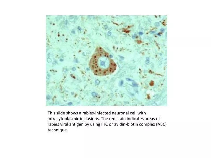
This slide shows a rabies-infected neuronal cell with intracytoplasmic inclusions. The red stain indicates areas of rabies viral antigen by using IHC or avidin-biotin complex (ABC) technique.
Confocal microscopy of double immunofluorescent staining for RCA-1 (red) and HIV gp41 protein (green). At least one of perivascular RCA-1 stained macrophages is infected with HIV, as evidenced by co-localization of RCA-1 and gp41 (yellow).
Immunofluorescence assay (IFA): SARS-CoV-infected Vero cells incubated with patient serum (1:50 dilution) obtained 11 days after the onset of symptoms, showing cytoplasmatic fluorescence. (Source: Institute for Medical Virology, Director: W. Doerr)