1 / 56

Figure 12.0 Mitosis

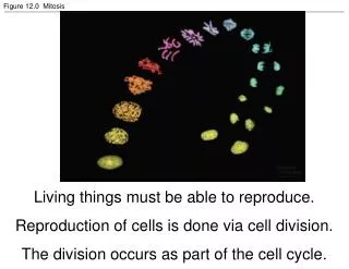
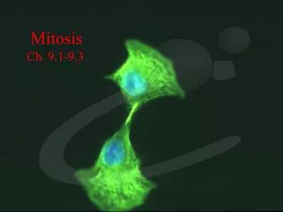
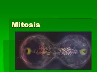
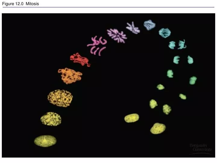
Figure 12.0 Mitosis. Figure 12.1a The functions of cell division: Reproduction. Figure 12.1b The functions of cell division: Growth and development. Figure 12.1c The functions of cell division: Tissue renewal. Figure 12.2 Eukaryotic chomosomes.

laksha
Download Presentation

Figure 12.0 Mitosis

An Image/Link below is provided (as is) to download presentation Download Policy: Content on the Website is provided to you AS IS for your information and personal use and may not be sold / licensed / shared on other websites without getting consent from its author. Content is provided to you AS IS for your information and personal use only. Download presentation by click this link. While downloading, if for some reason you are not able to download a presentation, the publisher may have deleted the file from their server. During download, if you can't get a presentation, the file might be deleted by the publisher.

E N D

Presentation Transcript


  1. Figure 12.0 Mitosis

  2. Figure 12.1a The functions of cell division: Reproduction

  3. Figure 12.1b The functions of cell division: Growth and development

  4. Figure 12.1c The functions of cell division: Tissue renewal

  5. Figure 12.2 Eukaryotic chomosomes

  6. Figure 19.0 Chromatin in a developing salamander ovum

  7. Figure 19.x1a Chromatin

  8. Figure 19.x1b Chromatin, detail

  9. Figure 12.3 Chromosome duplication and distribution during mitosis

  10. Figure 19.1 Levels of chromatin packing

  11. Figure 12.4 The cell cycle

  12. Figure 12.5 The stages of mitotic cell division in an animal cell: G2 phase; prophase; prometaphase

  13. Figure 12.5 The stages of mitotic cell division in an animal cell: metaphase; anaphase; telophase and cytokinesis.

  14. Figure 12.6 The mitotic spindle at metaphase

  15. Figure 12.7 Testing a hypothesis for chromosome migration during anaphase

  16. Figure 12.8 Cytokinesis in animal and plant cells

  17. Figure 12.5x Mitosis

  18. Figure 12.9 Mitosis in a plant cell

  19. Figure 12-09x Mitosis in an onion root

  20. Figure 12.10 Bacterial cell division (binary fission) (Layer 1)

  21. Figure 12.10 Bacterial cell division (binary fission) (Layer 2)

  22. Figure 12.10 Bacterial cell division (binary fission) (Layer 3)

  23. Figure 12.11 A hypothesis for the evolution of mitosis

  24. Figure 12.12 Evidence for cytoplasmic chemical signals in cell cycle regulation

  25. Figure 12.13 Mechanical analogy for the cell cycle control system

  26. Figure 12.14 Molecular control of the cell cycle at the G2 checkpoint

  27. Figure 12.15 The effect of a growth factor on cell division

  28. Figure 12.15x Fibroblast growth

  29. Figure 12.16 Density-dependent inhibition of cell division

  30. Figure 12.17 The growth and metastasis of a malignant breast tumor

  31. Figure 12-17x1 Breast cancer cell

  32. Figure 12-17x2 Mammogram: normal (left) and cancerous (right)

  33. Figure 13.1 The asexual reproduction of a hydra

  34. Figure 13.2 Two families

  35. Figure 13.x1 SEM of sea urchin sperm fertilizing egg

  36. Figure 13.3 Preparation of a human karyotype (Layer 1)

  37. Figure 13.3 Preparation of a human karyotype (Layer 2)

  38. Figure 13.3 Preparation of a human karyotype (Layer 3)

  39. Figure 13.3 Preparation of a human karyotype (Layer 4)

  40. Figure 13.x2 Human female chromosomes shown by bright field G-banding

  41. Figure 13.x3 Human female karyotype shown by bright field G-banding of chromosomes

  42. Figure 13.x4 Human male chromosomes shown by bright field G-banding

  43. Figure 13.x5 Human male karyotype shown by bright field G-banding of chromosomes

  44. Figure 13.4 The human life cycle

  45. Figure 13.6 Overview of meiosis: how meiosis reduces chromosome number

  46. Figure 13.7 The stages of meiotic cell division: Meiosis I

  47. Figure 13.7 The stages of meiotic cell division: Meiosis II

  48. Figure 13.8 A comparison of mitosis and meiosis

  49. Figure 13.8 A comparison of mitosis and meiosis: summary

More Related