Download
1 / 25
260 likes | 453 Views
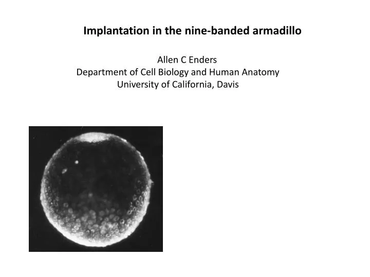
Implantation in the nine-banded armadillo. Allen C Enders Department of Cell Biology and Human Anatomy University of California, Davis.

E N D
Implantation in the nine-banded armadillo Allen C Enders Department of Cell Biology and Human Anatomy University of California, Davis
Implantation in the nine-banded (long-nosed) armadillo has several intriguing features. Initially the single blastocyst is confined to a chamber in the fundus of the uterus simplex during a prolonged period of delay of implantation. At implantation, trophoblast adjacent to the inner cell mass intrudes into pre-existing endometrial blood sinuses, which are subsequently exploited as the intervillous space of a villous hemomonochorial placenta. The endodermal layer of the blastocyst never extends much beyond the inner cell mass. Consequently, when at implantation the mural trophoblast disintegrates, the yolk sac is immediately inverted. The exocelom forms and expands precociously, and only as it expands is the epiblast divided into the four embryonic shields that constitute the origins of the identical quadruplet fetuses.
Zona-free blastocyst in delayed implantation. Blastocyst at the beginning of delayed implantation. The zonapellucida is starting to disintegrate. Section through the inner cell mass of a delay blastocyst.
TEM of the inner cell mass region of a delay blastocyst. Note the microvilli on the polar trophoblast cells. Delay blastocyst showing a distinction between polar trophoblast cells and ICM cells, but not a distinct endodermal layer.
Although there are some differences in the abundant ICM cells, there is no distinct endodermal layer.
Blastocyst flushed at about the time of implantation. The ICM appears to be forming an amniotic cavity by cavitation. Some of the polar trophoblast cells below may be becoming syncytial. Fundic endometrium during the delay period. Note the short glands and the large venous sinuses that are close to the endometrial surface.
Surface view of an implanting blastocyst after the uterus has been opened to expose the fundic chamber. The same blastocyst sectioned. Note that the blastocyst lumen has collapsed but that there is a large amnion and a couple of vesicle beginning to form an exocelom. There is a single epiblast. See labeling in the next slide.
At implantation, the mural (abembryonic) trophoblast is complete. Both the amnion and the exocelom form rapidly. Note that there is only a small stretch of parietal endoderm. A depiction of the relationship of the blastocyst to the endometrium at early implantation.
Plastic-embedded implantation site. The mesothelial lining of the exocelom is apparent. The trophoblast giant cells that have invaded the endometrium are not easily seen but occupy the pale area beneath the layer of cytotrophoblast that is backed by mesothelium. Note the large venous sinus on the lower left. A pale layer of mural trophoblast still covers the developing blastocyst.
Margin of an implantation site similar to that in the previous slide. A pale layer of mural trophoblast covers the visceral endoderm overlying the epiblast on the left, and the short stretch of parietal endoderm that abuts a circular mass of cytotrophoblast on the right. Pale trophoblast giant cells have invaded the endometrium and surround uterine glands. Note the layer of trophoblast cells overlying blood cells in the lower middle of the micrograph, and the mesothelial cells surrounding the exocelom.
The residual mural trophoblast is disintegrating. Note the polarity, as indicated by surface microvilli, of the endoderm overlying the inner cell mass.
Disintegration of the mural trophoblast inverts the implantation site, exposing the endoderm to the uterine lumen. Note the blood sinuses both below and above the endometrial glands. Plastic-embedded early-inverted implantation site. Although the epiblast layer is single, it is thicker towards the periphery. A cluster of cells at the margin of the implantation site constitutes the anchoring region to the endometrium. The pale trophoblast giant cells surround endometrial glands.
Margin of an implantation site showing the cytotrophoblast annulus that anchors to the endometrium. Both cytotrophoblast and pale giant cells extend beneath the uterine luminal epithelium {lower left}. Note troph remnant. Center of the previous site. Endoderm overlies epiblast. The amniotic epithelium is lined by mesothelial cells of the exocelom. Cuboidal mesothelial cells overlie a layer of cellular trophoblast, and giant cells overlie glands.
Diagram if the early inverted stage of implantation. Note that the uterine blood sinuses directly below the glands are continuous with the space between the giant cells and the layer of cellular trophoblast that borders the exocelom. The microvilli on this layer of trophoblast indicate its apparent absorptive nature.
Large pale giant trophoblast cells surround uterine glands. The cytotrophoblast layer above is backed by a squamous mesothelium . A venous sinus extends between regions where giant cells surround the glands, up to the layer of absorptive trophoblast at the top.
TEM of absorptive trophoblast. Note the abundant microvilli in the lower right. In this light micrograph the pale area over the tips of the layer of cellular trophoblast is composed of regions of microvilli. Note the close association of the trophoblastcells surrounding the dark gland cells below.
Later in implantation the surrounded gland cells begin to disintegrate. Eventually both the gland cells and trophoblast giant cells disintegrate, exposing the absorptive trophoblast to maternal blood sinuses and eliminating most of the fundic endometrium. TEM showing a gland cell with glycogen and lipid surrounded by a trophoblast giant cell.
While initially the mesothelial layer backing the absorptive trophoblast is simple, as in the above micrograph, eventually some fibers begin to appear and blood vessels form in situ, preceding vascularization of the absorptive trophoblast. Note the maternal blood cells in the space underlying the absorptive trophoblast.
In the upper left two villi are beginning to form, extending from the absorptive trophoblast layer. At a higher magnification the structure of the forming villi can be seen. There is a very thin layer of syncytial trophoblast overlying the villi. At their tips are numerous cytotrophoblast cells. Beneath the cytotrophoblast are numerous mesenchymal cells. Thus at this stage they are secondary villi.
Only at this stage when villi are forming does the epiblast divide into four separate embryonic shields. At first the amnion, although reduced, is still interconnected between shields.
At the time when primitive streaks first form, the four embryonic shields are widely separated at the margins of a large exocelom.
One of the four embryos seen in the previous slide, showing that they are in a late primitive streak stage with some mesodermal cells spreading over the endoderm of the yolk sac. The squamous mesothelial cells backing the amniotic epithelium are from the preexisting extraembryonic mesoderm.
As the conceptus develops it intrudes into the uterine lumen, exposing the inverted yolk sac to the luminal contents. Note the relatively small size of the embryo in the upper left compared to the overall size of the conceptus. Here the developing villi can be seen lining the area filled with maternal blood that is within the developing conceptus. Note also the large blood sinuses in the endometrium below. It is these sinuses that will eventually be invaded by the forming villi.
Margin of a forming placenta Villi with their dark masses of cytotrophoblastat their tips are intruding into the blood sinuses under the thick endometrium of the body of the uterus. The inverted yolk sac is in the lumen above, and villi are in the sinus below. The annulus is now a very thin area of attachment of trophoblast and yolk sac to the endometrium.
Publications Enders, A.C. 1960 Development and structure of the villous hemochorial placenta of the nine-banded armadillo. Journal of Anatomy 94:34-45. Enders, A.C. 1963 Fine structural studies of implantation in the armadillo. In A.C. Enders (Ed.), Delayed Implantation, University of Chicago Press, Chicago. Enders, A.C. 2002 Implantation in the nine-banded armadillo: how does a single blastocyst form four embryos? Placenta 23: 71-85. Enders, A.C. 2008 Placentation in armadillos, with emphasis on development of the placenta in polyembryonic species. Pp 172-180. In: Biology of the Xenarthra. W..J. Loughry and S.F. Vizcaino, eds., University Press of Florida, Gainesville.