Download
1 / 38
480 likes | 1.01k Views
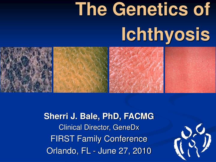
The Genetics of Ichthyosis. Sherri J. Bale, PhD, FACMG Clinical Director, GeneDx FIRST Family Conference Orlando, FL - June 27, 2010. What we’re going to talk about

E N D
The Genetics of Ichthyosis Sherri J. Bale, PhD, FACMG Clinical Director, GeneDx FIRST Family Conference Orlando, FL - June 27, 2010
What we’re going to talk about A primer on how ichthyosis genetically occurs, the chances of passing it along and what genetic tests are available and how they are administered
How many different ichthyoses are there? • > 20 disorders fit the definition of ichthyosis • > 10 other related disorders with more localized scaling/hyperkeratosis
How are ichthyoses classified? • Clinical features • Non-syndromic ichthyoses • Syndromic ichthyoses • Related disorders • Inheritance pattern • Gene defects • Etiology • Enzyme deficiencies • Structural protein defects • Regulatory protein defects • Other
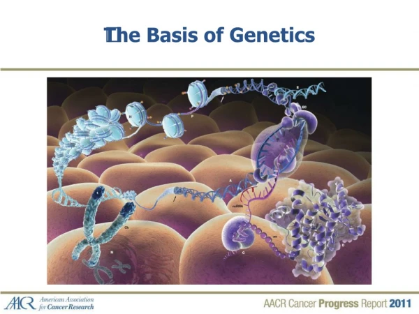
Genetics 101 • Chromosomes – structures inside the nucleus (command center) of the cell. • On the chromosomes, we carry genes • Genes are made up of a chemical called DNA • Chromosomes, and thus genes, are passed from parent to child following “rules of inheritance” [Mendel’s laws]
Types of Inheritance • X-linked • Recessive • Dominant (rare) • Autosomal • Recessive • Dominant
Steroid-Sulfatase Deficiency • X-linked recessive • Incidence 1:6000 males • Primary features • Onset between 1 and 3 weeks of age • Dark scale, tightly adherent • Most prominent on flexure surfaces (aka “Dirty neck” ichthyosis) • Asymptomatic corneal opacities (10-50%) • Cryptorchism (12-25%), increased risk of testicular cancer • The disease does not improve with age
Steroid-Sulfatase Deficiency • Diagnostic • plasma cholesterol sulfate levels • Assay to directly measure activity of steroid sulfatase is rarely done • Decreased placental sulfatase causes delayed onset/progression of labor in affected male fetuses • Genetics • STS gene on chromosome Xp22.32 • 90% of affected males have large intragenic deletions, or contiguous gene deletions
Autosomal Dominant Ichthyoses • Ichthyosis Gene Epidermolytic hyperkeratosis KRT1; KRT10 Epidermolysis Bullosa Siemens KRT2e Pachyonychia congenita I,II KRT6a,b, KRT16, KRT17 Epidermolytic PPK KRT9 Non-epidermolytic PPK many genes Keratitis-Ichth-Deafness (KID) GJB2 (GJB6) Erythrokeratoderma variabilis GJB3, GJB4
Epidermolytic Hyperkeratosis • Autosomal Dominant (1/2 the cases are due to new mutations) • Incidence 1:200,000-1:300,000 • Primary Features • Neonatal blistering, erosions and denuded skin • Progressive Hyperkeratosis, esp. of the flexures • Variable palm/sole involvement
Epidermolytic Hyperkeratosis • Genetics • Due to mutation in keratin-1 (KRT1) or keratin-10 (KRT10) gene • >40 different mutations, most are missense changes • >80% cluster at hot spots at the beginning or end of the gene • In 30% of all EHK patientsmutations occur at Arg156 in KRT10
? How can you say its autosomal dominant? I’m the only person in my family with this disorder!
Ovaries Testes Egg cell Sperm Mutation Mutation Germline Mutation
Conception Mutation Disease Germline Mutation
I have a been diagnosed with an ‘Epidermal Nevus’. What is it and how does it come about? ?
Zygote Mutation Post-zygotic mutation Gametes Two cell lineages Mosaic Cell Migration ‘Epidermal Nevus’ = Skin Mosaicism for Mutation
Mosaicism • Due to DNA Mutation that occurs during mitosis of a single cell at early stages of fetal development • “post-zygotic mutation” • All descendent cells will carry the mutation, other cells are normal • Gives rise to two (or more) genetically distinct cell lines derived from a single zygote • Mosaicism can affect somatic and/or germline tissues • Generally only parts of the organism are affected
? I have an ‘Epidermal Nevus’. Should I be worried about my children? • If germline is affected, mutation can be transmitted to the offspring resulting in full-blown disease
? What is my risk of having an affected child? • Greater than the population risk for a new mutation • Depends on what percentage of germ cells harbor mutation • Rule of thumb: • Small nevus-- • small risk • Large nevus on both sides of the body--high risk
Autosomal Recessive Ichthyoses • Ichthyosis Gene Harlequin ichthyosis ABCA12 • Lamellar ichthyosis TGM1, ABCA12 • CIE ALOXE3; ALOX12B Ichthyin • Cytochrome P450 • Sjögren-Larsson syndrome ALDH3A2 • Neutral lipid storage disease CGI-58 (ABHD5) • Netherton syndrome SPINK5 • Refsum disease PAHX, PEX7 • Trichothiodystrophy+Ichthyosis ERCC2; ERCC3
Lamellar Ichthyosis • Autosomal Recessive • Incidence 1:200,000 • Primary Features: • Collodion baby phenotype • Plate-like, large, dark scale • Ectropion, Eclabium • Scarring alopecia
Lamellar Ichthyosis • Due to mutation in the TGM1 gene in the vast majority of cases, coding for Transglutaminase-1 • A few common mutations exist (the “German” splice-site mutation) and R141 and R142 in exon 3. • Few families with mutation in ABCA12, Ichthyin, and cytochrome P450 genes
Congenital Ichthyosiform Erythroderma • Autosomal Recessive • Incidence 1:200,000-300,000 • Primary features • Collodion baby presentation • Bright red (erythrodermic) skin • Fine, white scale
Congenital Ichthyosiform Erythroderma • Due to mutation in many different genes, 5 of which are known • ALOX12B and ALOXE3 (in about ~10%) • Ichthyin (in about ~10%) • A new cytochrome P450 gene • Enzymes encoded by these genesare involved in lipid metabolism • Operate in common membrane arachidonic acid pathway (lipoxygenase)
Harlequin Ichthyosis • Autosomal recessive • Mutation in ABCA12 gene • (ATP-binding cassette transporter protein) • Primary features: • Thick, armor-like plates of scale that fissure and crack • Eclabium and Ectropion • Poor prognosis, although survivors have a congenital ichthyosiform erythroderma phenotype
How do we detect a mutation? • Karyotype • arrayCGH • FISH • Chromosomes • DNA • Metabolic • Sequence analysis • Mutation scanning • Targeted mutation analysis • Analytes • Enzyme assay
What do we need for mutation testing? • Material required for testing: • Buccal swabs • Blood • Skin punch biopsy
Gly278Arg How is DNA Sequencing Done?
What is the use of this mutation information ? • Identification of disease-causing mutation(s) allows answers to the questions: • What do I have? • Why do I have it or how did it happen? • What is the chance it will happen again? • What’s wrong with my skin and how best can it be treated?