Download
1 / 20
210 likes | 434 Views
FRACTURES OF THE PROXIMAL HUMERUS. Presented by Mahsa Mehdizade Dr. Mardani Porsina Hospital Spring 1392. Incidence. Proximal humerus fxs comprise 4-5% of all fxs. Minimal displacement 80% Two-part fxs 10% Three-part fxs 3% Four-part fxs 4% Articular surface fxs 3%. Anatomy.

E N D
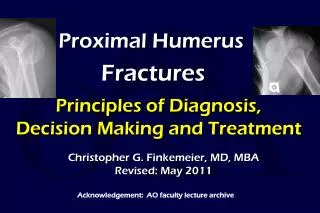
FRACTURES OF THE PROXIMAL HUMERUS Presented by Mahsa Mehdizade Dr. Mardani Porsina Hospital Spring 1392
Incidence • Proximal humerus fxs comprise 4-5% of all fxs. • Minimal displacement 80% • Two-part fxs 10% • Three-part fxs 3% • Four-part fxs 4% • Articular surface fxs 3%
Anatomy • Comprised of four segments: • Humeral head • Greater tuberosity • Lesser tuberosity • Humeral shaft
Neurovascular Supply • Anterior and posterior humeral circumflex arteries • Arcuate artery-continuation of the ant humeral circumflex and supplies most of the humeral head. • Axillary nerve-most commonly injured
Forces on Segments • Greater tuberosity is displaced superiorly and posteriorly by the supraspinatus and external rotators. • Lesser tuberosity is displaced medially by the subscapularis. • The shaft is displaced medially by the pectoralis major.
Mechanism of Injury • Elderly, osteoporotic, usually female: fall on outstretched arm. • Young adults: high-energy trauma; usually more severe fxs and dislocations
Radiographic Evaluation • A/P view • Scapular Y view • Axillary view • Best view for glenoid articular fxs and dislocations • CT scan: helpful in evaluating articular involvement and degree of displacement
Classifications • Neer-four parts: greater and lesser tuberosities; shaft; humeral head. • A part is displaced only if >1cm of displacement or 45 degrees of angulation is present. • At least 2 views must be obtained • AO-emphasizes the vascular supply to the articular segment • Three types: • Type A: Extraarticular unifocal fxs • Type B: Extraarticular bifocal fxs • Type C: Articular fxs • Not commonly used
Closed reduction Immobilization Early ROM if stable External stabilization Percutaneous pins External fixator Ilizarov frame Open reduction and internal fixation Screw fixation Tension banding Buttress plating Fix-Clip system Intramedullary fixation Rush rods Ender’s nails Nails with interlocking screws Excisional arthroplasty Hemiarthroplasty Treatment Options
Fractures to Consider for Closed Treatment Minimally displaced 2 part fx’s (or positional reduction of significant displacement) GT fractures should be <5mm). Minimally displaced 3- and 4-part fractures
Fractures to Consider for ORIF • Displaced GT fx (> 5 mm) • LT fx with involvement of articular surface • Displaced or unstable surgical neck fx • Displaced anatomic neck fx in young pt. • Displaced, reconstructible 3- and 4-part fractures
Fractures to Consider Hemiarthroplasty • Young/Middle age • nonreconstructable articular surface (severe head split) or extruded anatomic neck • Elderly • many 4 parts • some severe 3 parts • most 3,4 part fracture dislocations • most head splits
Potential Complications • Neurologic injury • Brachial plexus-Stableforth reported an incidence of 6.1% • Axillary-common • Vascular injury • Stableforth also reported a 4.9% incidence of arterial injury with displaced fxs; most commonly the axillary artery • An intact radial pulse doe not exclude an arterial injury so keep it in mind.
Complications cont. • Avascular necrosis • Hagg and Lungberg reported an incidence of 3 – 14% with 3- part fxs and 13 – 34% with 4-part fxs, using closed reduction. • Nonunion (uncommon) • Malunion – often associated with AVN • Adhesive capsulitis • Myositis ossificans • Infection