Download
1 / 34
350 likes | 496 Views
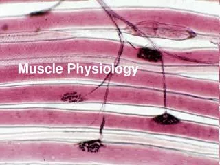
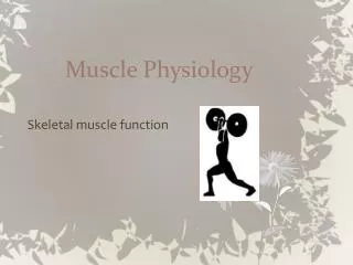
Muscle Physiology. Chapter 12. Muscle Tissue. Specially designed to contract Generates mechanical force Functions locomotion and external movements internal movement (circulation, digestion) heat generation. Types of Muscle. Skeletal - attached to skeleton

E N D
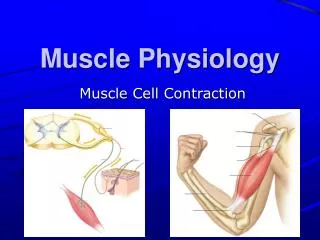
Muscle Physiology Chapter 12
Muscle Tissue • Specially designed to contract • Generates mechanical force • Functions • locomotion and external movements • internal movement (circulation, digestion) • heat generation
Types of Muscle • Skeletal - attached to skeleton • Smooth - found in walls of hollow visceral organs • Cardiac - in heart
Skeletal Muscle • Connected to skeleton via tendons • attached to immobile bone at one end (origin) • other end attached to more moveable bone (insertion) • Many muscles are used to bend the skeleton at joints • Antagonistic pairs • Contraction of one does the opposite of its counterpart
Skeletal Muscle Organization • Muscle fibers (cells) • elongate cells, parallel arrangement, • bundled by connective tissue • multiple nuclei • striated • sarcolemma – cell membrane • sarcoplasmic reticulum (SR) • Internal membraneous network • myofibrils - intracellular contractile elements • thick filaments • thin filaments Figs 12.1, 12.5, & 12.15
Myofibril Structure • Composed of sarcomeres • smallest functional unit of muscle • repeating units of thin and thick protein filaments • Thick Filament = Myosin • Thin Filament = Actin, Troponin, Tropomyosin Figs 12.6 & 12.7
Thick Filament Structure • Bundles of several hundred myosin molecules • intertwining tails + globular heads • heads contain: • actin binding sites • ATP-hydrolyzing sites • project outward towards actin • form crossbridges • bonds with actin • Important during contraction Fig 12.10
Thin Filament Structure • Actin • primary structural protein • spherical protein subunits connected in long, double strand • Contains myosin binding site • Tropomyosin • threadlike proteins • normally cover myosin binding sites • Troponin • Ca2+ Binding Protein • holds tropomyosin in place Figs 12.10, 12.13
Thin Filament Structure • When Ca2+ binds to troponin • Shape of troponin changes • Shifts tropomyosin off myosin binding sites • Myosin binds to actin Fig 12.14
Skeletal Muscle Contraction:Sliding Filament Mechanism • Movement of thin filaments over thick • sarcomere shortening • thick filaments are stationary; thin are dragged across thick • length of the filaments do not change. Fig 12.9
Crossbridge Cycling • Myosin head binds to actin • Cross bridge bends (Power Stroke) • thin filaments pulled toward center of sarcomere • Cross bridge link broken • Cross bridge ‘unbends’ and binds to next actin molecule Figs 12.11, 12.12
Muscle Contraction • ATP required • ATP must bind to myosin for myosin to release actin • Ca2+ required • binding of Ca2+ to troponin uncovers myosin binding sites on the actin • Ca2+ released inside the cell to induce contraction
Neural Activation of Muscle Contraction • Somatic Motor Neurons stimulate AP’s in skeletal muscle cells (neurotransmitter generates EPSPs) • Excitation-Contraction Coupling • Events that link muscle excitation (action potential) to muscle contraction (cross-bridge cycling) • Triggers release of Ca2+ into the cytosol of the muscle fiber
Neural Input • Somatic motor neurons • under voluntary and involuntary control • controlled by interneurons in motor cortex and cerebellum Fig 9.1
Neural Input • Neuromuscular Junction (NMJ) • synapse between motor neuron and muscle fiber • Presynaptic Terminal • enlarged axon terminal • contains acetylcholine (ACh) • Motor End Plate • Subsynaptic membrane • Chemically-gated Na+ channels • Acetylcholinesterase enzyme • breaks down ACh Figs 12.3, 12.16
Sequence of Events • AP travels down axon to terminal • Exocytosis of ACh • ACh diffuses across cleft • ACh binds to ACh-gated Na+ channels • Opens ion channels • Graded depolarization AP in sarcolemma of muscle fiber • AP results in release of Ca2+ inside muscle fiber • Muscle contracts and shortens Fig 12.16 http://www.blackwellpublishing.com/matthews/nmj.html
Excitation-Contraction Coupling • Sarcoplasmic Reticulum (SR) • modified ER • surrounds myofibrils • stores Ca2+ • Linked to the sarcolemma by transverse tubules Fig 12.15
Excitation-Contraction Coupling • T-tubules conduct signal from APs into the cell • voltage-gated Ca2+ channels open • Ca2+ released into cytosol • Binds to troponin • Troponin moves tropomyosin • Crossbridge formation ensues Figs 12.14, 12.15 http://www.blackwellpublising.com/matthews/myosin.html
Muscle Relaxation • Ca2+ pumped back into the SR by active carrier-mediated transport • troponin releases Ca2+ • tropomyosin covers myosin binding sites on the actin molecules • Membrane-bound enzyme (acetylcholinesterase) • breaks down ACh released at the NMJ
All or None • Individual muscle fibers respond to a single stimulus in an all or none fashion • undergo action potential • action potential triggers contraction • stimulus < threshold = no contraction • stimulus > threshold = maximal contraction
Motor Units • Multiple muscle fibers are enervated by a single motor neuron • Motor Unit • motor neuron + all muscle fibers it innervates • muscle fibers in a motor unit contract as a single unit Fig 12.4
Motor Unit Recruitment • Individual motor units contract in an all-or-none fashion • Differences in contractile strength are due to differences in the number of contracting motor units • Motor Unit Recruitment • increasing the number of contracting motor units to increase the overall strength of contraction
Motor Units and Control of Movement • Different regions of the body have different numbers and sizes of motor units • Leg muscles • strong contractions, little precision of movement • large motor units (2000 fibers/unit) • few individual units • Finger muscles • weaker contractions, more precise movements • small motor units (10 fibers/unit) • many individual units
Motor Units and Control of Movement • More motor cortex area allocated to control of areas with more numerous, smaller motor units • Precise control of the strength of muscle contractions Fig 8.7
Energy for Muscle Activity • Muscle contraction requires ATP • cross bridge cycling • Ca2+ pump activity • Sources available • cytosolic ATP • creatine phosphate • aerobic respiration • anaerobic respiration
ATP and Creatine Phosphate • ATP in muscle • limited (used up in a few contractions) • Creatine phosphate • storage of high energy phosphate bonds • used to quickly regenerate ATP from ADP • limited supply in cells Fig 12.22
Aerobic Respiration • Occurs in mitochondria • Requires O2 to form ATP • fatty acids = primary nutrient source • contain lots of energy, but requires O2 to release it • O2 transported in by blood (hemoglobin) and also stored in muscle tissue (myoglobin) • Aerobic exercise • light to moderate exercise (walking, jogging, swimming) • Maximum oxygen uptake • max rate of O2 delivery to the muscles • max level of aerobic activity
Anaerobic Respiration • Glycolysis + lactate fermentation • breakdown of glucose • stored as glycogen in muscle cells • Does not require O2 • generates ATP quickly (faster than aerobic respiration) • Used during intense exercise • anaerobic exercise • O2 supply cannot keep up with demand • Lactate produced • muscle soreness and fatigue Fig 12.21
Consequences of Anaerobic Respiration • Muscle Fatigue • inability to maintain tension due to previous contractile activity • ATP stores used up • ion gradients across membrane disrupted • high lactate levels inhibit contractile protein function • Oxygen Debt • increased O2 consumption (breathing) after exercise • restore myoglobin and hemoglobin O2 content, metabolize lactate, etc.
Changes with Regular Sustained Exercise • number and size of mitochondria • number of blood capillaries supplying muscle • O2 and nutrients + more efficient waste removal • amt. myoglobin in muscle tissue • size of muscle fibers (weight training) • # of myofibrils • no change in # of muscle fibers
Smooth Muscle • visceral organs • small tapered fibers • single nuclei • lack striations • poorly developed sarcoplasmic reticulum Fig 12.33
Smooth Muscle Contraction • No striations • contractile proteins not arranged in sarcomeres • arranged in fish-net network • allows for extensive contraction, even when stretched Fig 12.33
Smooth Muscle Excitation-Contraction Coupling • Depolarization of sarcolemma opens voltage-gated Ca2+ channels • can open in response to graded potentials • contraction strength is proportional to stimulus strength • Ca2+ enters the cell from the extracellular fluid
Smooth Muscle Excitation-Contraction Coupling • Ca2+ binds with calmodulin in cytoplasm • Ca2+-calmodulin binds to myosin light chain kinase (MLCK) • activates MLCK • MLCK phosphorylates myosin • needed for myosin to bind actin • Cross-bridge cycling Fig 12.34