Download
1 / 57
660 likes | 1.3k Views
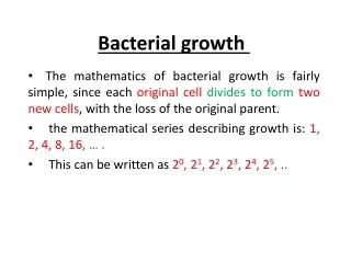
Bacterial Exotoxins. First bacterial virulence factors identified Diphtheria toxin isolated in 1888.

E N D
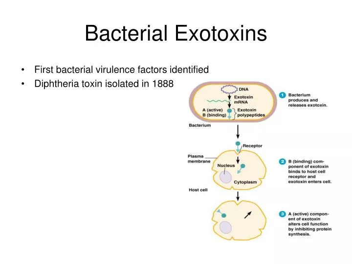
Bacterial Exotoxins • First bacterial virulence factors identified • Diphtheria toxin isolated in 1888
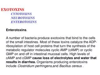
Enterocyte intracellular signaling leading to intestinal secretion. Four main pathways seem to be involved in intestinal secretion of water and electrolytes: cAMP, cGMP, Ca, and cytoskeleton. These pathways are activated by several enteric pathogens, either directly or through elaboration of enterotoxic products. CT, cholera toxin; LT, heat-labile enterotoxin; TDH, thermostable direct hemolysin; CD, Clostridium difficile; EAST1, enteroaggregative E. coli heat-stable toxin 1; STa, heat-stable toxin a; AC, adenylate cyclase; GC, guanylate cyclase; CM, calmodulin; PKC, protein kinase C; ZOT, zonula occludens toxin; EGF-R, epidermal growth factor receptor; ECM, extracellular matrix; MAPK, mitogen-activated protein kinase.
Mode of action of CT. A: adenylate cyclase, located in basolateral membrane of intestinal epithelial cells, is regulated by G proteins. CT binds via the B subunit pentamer to GM1 ganglioside receptor inserted in lipid bilayer.
the A subunit enters the cell, perhaps via endosomes, and is proteolitically cleaved into A1 and A2 peptides. A1 is activated and transfers an ADP-ribose moiety (ADPR) and NAD to the -subunit of Gs protein. ADP-ribosylated -subunit dissociates from other subunits of Gs and activates adenylate cyclase, thereby increasing intracellular cAMP concentration. Three possible scenarios have been proposed to explain entry of the toxin and activation of adenylate cyclase: 1) A1 subunit translocates through apical membrane, leaving B pentamer on apical membrane; 2) A1 peptide ADP-ribosylates an -subunit in apical membrane, and the ADP-ribosylated -subunit traverses the cell to attach to adenylate cyclase located in basolateral membrane; 3) entire toxin enters cell via endosomes, and A subunit translocates through endosomal membrane, then A1 peptide ADP-ribosylates Gs located in basolateral membrane, perhaps after endosome-plasma membrane fusion. ARF, adenosine rybosil factor.
increased cAMP activates protein kinase A, leading to protein phosphorylation. Protein phosphorylation leads to increased Cl ion secretion in crypt cells and decreased NaCl-coupled absorption in villus cells.
Exotoxins • They are toxic proteins secreted from a living bacterium. They alter the normal metabolism of host cells and have deleterious effects on the host. • They are different from ENDOTOXINS (LPS or LOS). • Exotoxins are diverse i.e., they have different structures and different modes of action. • Some will form pores in the host cell plasma membrane, others will bind receptors, and others will be internalized by the host cell. • One pathogen may produce one toxin or one pathogen may produce several toxins • S. aureus produces 25 or 30 different toxins.
Toxin Classification • Type I toxins: superantigens • They bind to the host cell surface, but they are not translocated into the cell • They will modulate the immune response
Toxin Classification • Type II toxins: pore forming toxins • They act on the host cell membrane. • Host cells will leak then die
Toxin Classification • Type III toxins: A-B toxins • They bind to the host cell one specific receptor and are translocated into the cell. • They will become active and modify some proteins or other components of the host cell.
A-B toxins Cell surface Active Binding A B
Toxin Classification Effector proteins that are translocated into host cells through a type III secretion system. This type is present in many gram negative bacteria. No binding, direct injection.
Toxin effects • Toxins that lyse cells (hemolysins, leukocidins) • Toxins that elevate cyclic AMP (cholera toxin) • Toxins that block protein synthesis (diphtheria toxin) • Toxins that block nerve function (botulinum, tetanus)
Domain Organization • A domain; catalytic domain. • B domain; receptor-binding domain. • AB5 toxins; consist of 6 non-covalently bound proteins.
Domain Organization • AB toxins; one single protein. • Diphtheria has an N-terminal catalytic domain and a C-terminal domain receptor-binding domain. • A-B toxins; proteins that are not associated in solution, but associate upon binding to the host cell. • Anthrax toxins (edema toxin and lethal toxin). • This type is synthesized as two different proteins.
What is the Role of a Toxin In Disease? • Toxins may have several roles (these are not mutually exclusive) • Directly toxic to host • Aid in the establishment of the disease • help in the acquisition of nutrients • interfere with the normal function of immune, or other cell types, cells during the infection • The contribution of toxins to disease remains unclear or incompletely understood
Diphtheria • Diphtheria is normally an infection of the throat caused by a bacterium, Corynebacterium diphtheriae. • Gram-positive • Non-spore forming • Non-motile • Aerobic rod • The tox gene codes for the AB toxin.
Pathogenesis of Diphtheria • Encounter – Corynebacterium diphtheriae encountered only from other people (carriers) • Entry – respiratory droplets; organism colonizes pharynx • Spread • Multiplication – iron likely a factor • Evasion of host immune response – adhesins; toxin may kill phagocytes contributing to pseudomembrane • Damage – inflammation; circulating toxin • Transmission – aerosolized droplets;
HPR10 • Diphtheria: infection of upper respiratory tract by Corynebacterium diphtheriae • bacteria grow on throat tissues • characteristic formation of pseudomembrane (greyish membrane of bacteria, damaged host cells) as a result of host’s inflammatory response • systemic exotoxin release is responsible for tissue damage • tox gene carried by lysogenic bacteriophage → only lysogenized bacteria cause serious disease • formerly major childhood disease; now rare due to DTP vaccine • “D” component of vaccine is formalin-treated diphtheria toxin (= toxoid) • a toxoid is nontoxic but remains immunogenic pseudomembrane • other A-B toxins include: • the neurotoxin of Clostridium botulinum • tetanus toxin (neurotoxin produced by Clostridium tetani) • pertussis toxin (whooping cough, Bordetella pertussis) • Shiga toxin (bacterial dysentery, Shigella dysenteriae)
tox Gene • There are toxigenic and nontoxigenic strains of Corynebacterium diphtheriae. • The tox gene encoding DT is carried by a family of corynebacteriophages • Toxigenic strains of Corynebacterium diphtheriae are lysogenized by these phages.
Catalytic Region A Subunit B Subunit Translocation Region Receptor-Binding Region Molecular Structure of Diphtheria Toxin
Domain Structure of Dt • R domain or C-terminal receptor-binding (R) domain: It binds to cell surface receptor, allowing the toxin to enter the cell by receptor-mediated endocytosis. • C domain or N-terminal catalytic (C) domain: It blocks protein synthesis by transfer of ADP-ribose from NAD to a diphthamide residue of EF-2. • T domain or central domain of diphtheria toxin is the translocation (T) domain: pH-induced conformational change of this domain triggers insertion into the endosomal membrane and facilitates the transfer of the catalytic domain into the cytoplasm.
Binding to the host-cell receptor: • The R-domain, which is part of the B-chain, binds to a specific receptor on the host cell surface. • This receptor is the heparin-binding epidermal growth factor (HB-EGF) precursor. • This is a membrane protein expressed on the host cell.
Endocytosis and Translocation • Endocytosis of the toxin-receptor complex. • Acidic pH in the endosome promotes a conformational change (T-domain) that inserts the toxin into the vesicle membrane. • The A-chain is translocated into the cytoplasm. • Reduction of the disulfide releases the A-chain into the cytoplasm.
Enzymatic activity • In the cytoplasm, the A-chain catalyzes the ADP-ribosylation of EF-2. • ADP-ribosylation uses NAD as a substrate. • In eukaryotes, EF-2 participates in the elongation step of translation. • ADP-ribosylation renders EF-2 inactive and blocks protein synthesis. • Causes cell to die. • ADP-ribosylation occurs at an unusual derivative of histidine, called diphtamide (post-translational modification). • Physiological function of diphtamide is unknown. • A single molecule of A-chain is enough to kill one eukaryotic cell. • Very Powerful.
Diphtheria Vaccine • Toxoid: the toxin is rendered nontoxic but still immunogenic by chemical modification or heat treatment. • Diphtheria toxoid = Toxin treated with formaldehyde is nontoxic but immunogenic. • Formaldehyde reacts with Lys amino groups and forms internal cross-links [prot]-NH-CH2-NH-[prot]
Immune Status to C. diphtheriae and Sensitivity to Diphtheria Toxoid TOXIN TOXOID Diagnostic Schick Skin Test
Anthrax toxin is an AB-type toxin “A” moiety is the effector molecule • “B” is the receptor-binding domain which facilitates the “A” moiety’s entry into the host cell • Two different types of “A” moieties -- EF and LF • “B” moiety is PA • EF and LF moieties cannot produce pathogenic effect on their own -- they must be present with PA moiety
Edema factor • Edema factor (EF) is called what it is due to its effect in producing edema in animals injected with it • EF is a calmodulin-dependent adenyl cyclase enzyme -- increases the concentration of cAMP in the cells • This increase in cAMP upsets water homeostasis in cells -- this may be the cause of the edema observed • 84 kDa