Download
1 / 16
160 likes | 268 Views
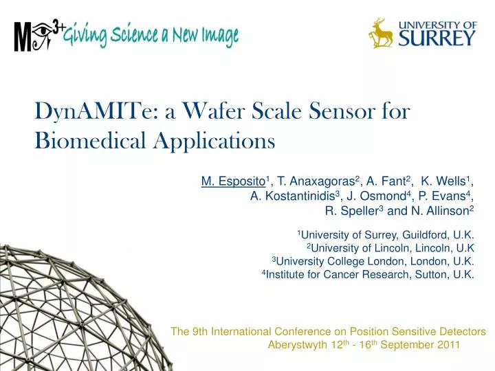
+. DynAMITe: a Wafer Scale Sensor for Biomedical Applications. M. Esposito 1 , T. Anaxagoras 2 , A. Fant 2 , K. Wells 1 , A. Kostantinidis 3 , J. Osmond 4 , P. Evans 4 , R. Speller 3 and N. Allinson 2. 1 University of Surrey, Guildford, U.K. 2 University of Lincoln, Lincoln, U.K

E N D
+ DynAMITe: a Wafer Scale Sensor for Biomedical Applications M. Esposito1, T. Anaxagoras2, A. Fant2, K. Wells1, A. Kostantinidis3, J. Osmond4, P. Evans4, R. Speller3 and N. Allinson2 1University of Surrey, Guildford, U.K. 2University of Lincoln, Lincoln, U.K 3University College London, London, U.K. 4Institute for Cancer Research, Sutton, U.K. The 9th International Conference on Position Sensitive Detectors Aberystwyth 12th - 16th September 2011
1/14 Contents • Biomedical imaging requirements • Large area imaging detector with bimodal dynamic range and resolution • Pixel structure and readout architecture • Optical testing • Conversion gain/ Read Noise/ Quantum efficiency/ FWC • Charge collection test • Non destructive readout- two separated readouts M.Esposito PSD9 Aberystwyth 15.09.2011
2/14 Large area imaging detectors Large Area Detectors Near real time frame rate K. Lee et al., J. Bone Joint. Surg. Am. 2010;92:2709-18. http://lemur.cmp.uea.ac.uk/ • Flat Panel Imagers • Large pixels • High noise • Low frame rate • Artefacts CMOS APS http://www.aesociety.org/ M.Esposito PSD9 Aberystwyth 15.09.2011
3/14 Biomedical Imaging detectors Low Noise High Spatial Resolution High Dynamic range Large Pixels → High dynamic range Small Pixels → Low Noise High Spatial Resolution M.Esposito PSD9 Aberystwyth 15.09.2011
4/14 The DynAMITe detector 12.8 cm × 12.8 cm 2 side buttable Dynamic range Adjustable for Medical Imaging Technology M.Esposito PSD9 Aberystwyth 15.09.2011
5/14 DynAMITe: sensor architecture 100 mm 1260×1280 Pixels (P) 100mm pitch 50 mm 2520×2560 Sub-Pixels (SP) 50mm pitch 12.8 cm Different reset voltages → Different depletion widths 12.8 cm M.Esposito PSD9 Aberystwyth 15.09.2011
6/14 Single Pixel readout Select (S)P1 Reset (S)P1 Pixel Reset (S)P2 • Frame rate • SPi @15 fps → SP @30 fps • Pi @45 fps → P @90 fps • Region of interest (100×100) • SPi @370fps → SPi @740 fps Select (S)P2 Out (S)P2 Out (S)P1 M.Esposito PSD9 Aberystwyth 15.09.2011
7/14 DynAMITe: readout Pixels Sub-Pixels Pixels & Sub-Pixels Pixels & Sub-Pixels (ROI) Sub-Pixels & Pixels (ROI) Non destructive readout Online dose sensing M.Esposito PSD9 Aberystwyth 15.09.2011
8/14 Radiation hardness A1 B1 C1 D1 B2 C2 D2 A2 38 MeV protons 1.5 MeV photons M.Esposito PSD9 Aberystwyth 15.09.2011
9/14 First images with DynAMITe M.Esposito PSD9 Aberystwyth 15.09.2011
10/14 Optical performance (Sub-Pixels) 523 nm M.Esposito PSD9 Aberystwyth 15.09.2011
11/14 Charge collection tests M.Esposito PSD9 Aberystwyth 15.09.2011
12/14 Non destructive readout (I) Signal SP2 T2 SP2 2×T2 RESET 3×T2 4×T2 SP1 T2 2×T2 3×T2 4×T2 = T1 time SP2 sampled @ T2 SP1 sampled and reset @T1 T2<T1 M.Esposito PSD9 Aberystwyth 15.09.2011 ResetSP1
13/14 Non destructive readout ROI (SP2) sampled @ 26.3 fps Full frame (SP1)sampled and reset @3.6 fps M.Esposito PSD9 Aberystwyth 15.09.2011
14/14 Conclusions • Dynamite sensor • High dynamic range • Low Noise • High Spatial Resolution • Several readout modalities • Optical characterisation (Sub-Pixel camera) • 45% QE • 150 e- read noise • 2.8 × 10⁵ e- FWC • Charge collection tests / Non destructive readout • Future applications: • Radiotherapy portal imaging • Breast mammography • Diffraction imaging • Electrophoresis M.Esposito PSD9 Aberystwyth 15.09.2011