Download
1 / 24
240 likes | 398 Views
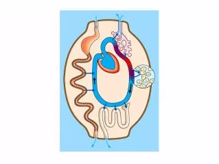
Respiration. Metabolism. Respiration. ostia. Semilunar valves. Left atrium. Right atrium. Atrioventricular valves. Left ventricle. Right ventricle. The Heart : Control - Cardiac cells : myogenic (self-excitable) in syncytium

E N D
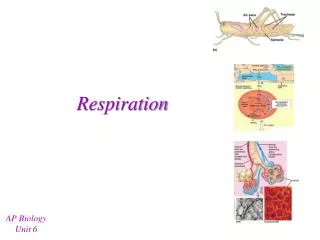
Respiration Metabolism
ostia Semilunar valves Left atrium Right atrium Atrioventricular valves Left ventricle Right ventricle
The Heart: Control -Cardiac cells: myogenic (self-excitable) in syncytium Controlled by sinoatrial (SA) node, the pacemaker. 1st: Atrial electrical wave -> two atria contract 2nd: Wave at atrioventricular (AV) node, delayed 0.1s 3rd: Common bundle & Purkinje fibers to Ventricles -Regulation of SA and AV nodes: Nervous: Sympathetic vs. Parasympathetic (vagus) Hormones: Epi, ACh, Temperature, Exercise, pH these also effect blood vessels (constrict or relax PR)
Sinoatrial node Left atrium Right atrium Atrioventricular node Left ventricle Right ventricle Conducting fibers
SA node activates atria AV node delay Electrical activity in atria Electrical activity in ventricles Ventricles recover 0 0.1 0.2 0.3 0.4 0.5 Time (seconds)
Fig. 42-9-1 1 Pacemaker generates wave of signals to contract. SA node (pacemaker) ECG
Fig. 42-9-2 2 Signals are delayed at AV node. AV node
Fig. 42-9-3 3 Signals pass to heart apex. Bundle branches Heart apex
Fig. 42-9-4 Signals spread throughout ventricles. 4 Purkinje fibers Be the HEART and Depolarize
The Heart: Function -Functionpump blood; create pressure gradient. Contraction and relaxation =cardiac cycle During systole, muscle contracts and pumps During diastole, muscle relaxes and chambers fill Sounds “Lub” AV, “Dub” SL, “ssssshh” murmur.
Blood returning to the mammalian heart in a pulmonary vein drains first into the vena cava. left atrium. right atrium. left ventricle. right ventricle.
Heart valves function to _____. • keep blood moving forward through the heart • mix blood thoroughly as it passes through the heart • control the amount of blood pumped by the heart • slow blood down as it passes through the heart • propel blood as it passes through the heart
Fig. 42-14 Direction of blood flow in vein (toward heart) Valve (open) Skeletal muscle Valve (closed)
Compared with the interstitial fluid that bathes active muscle cells, blood reaching these cells in arteries has a • higher PO2. • higher PCO2. • greater bicarbonate concentration. • lower pH. • lower osmotic pressure.
What is unique about blood in pulmonary arteries compared with blood in other arteries? • Blood in pulmonary arteries is always blue; it is red in all other arteries. • It is moving away from the heart. • It is moving toward the heart. • It is the same as blood in other arteries. • It is loaded with carbon dioxide.