Download
1 / 32
320 likes | 639 Views
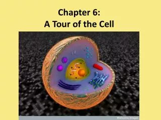
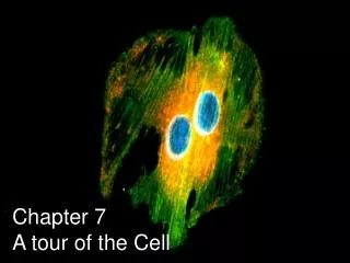
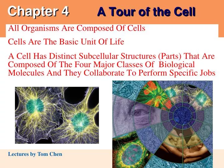
Chapter 4 A Tour of the Cell. 0. All Organisms Are Composed Of Cells Cells Are The Basic Unit Of Life A Cell Has Distinct Subcellular Structures (Parts) That Are Composed Of The Four Major Classes Of Biological Molecules And They Collaborate To Perform Specific Jobs .

E N D
Chapter 4 A Tour of the Cell 0 All Organisms Are Composed Of Cells Cells Are The Basic Unit Of Life A Cell Has Distinct Subcellular Structures (Parts) That Are Composed Of The Four Major Classes Of Biological Molecules And They Collaborate To Perform Specific Jobs
The Art of Looking at Cells • Early scientists who observed cells • Made detailed sketches of what they saw • You will do this in the Microscopes and Cells lab too. • These early sketches revealed an important relationship • Between art and biology, the most visual of the sciences
Eyepiece Ocularlens Objective lens Specimen Condenserlens Lightsource INTRODUCTION TO THE CELL • 4.1 Microscopes provide windows to the world of the cell • The light microscope (LM) • Enables us to see the overall shape and structure of a cell Figure 4.1A
Light microscopes Magnify cells, living and preserved, up to 1,000 times The LM is particularly good for looking at living cells and cells and tissues that have been stained TEM 2,800 SEM 2,000 Figure 4.1D Figure 4.1C • The electron microscope • Allows greater magnification and reveals cellular details Scanning electron microscopes (SEM) images are usually about 10,000–20,000 times larger than life size. The SEM is particularly good for showing organismal and cellular surfaces under high magnification Transmission electron microscopes (TEM) images are usually about 100,000–200,000 times larger than life size. The TEM is particularly useful for showing the internal structures of cells
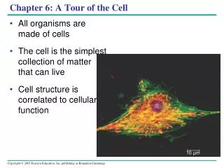
Overview: The Fundamental Units of Life • The cell is the basic unit of life. • All organisms are made of cells. • New cells are formed from other existing cells. • A cell is a fundamental unit of structure, function and organization in all living organisms.
4.2 Most cells are microscopic, Cells vary in size and shape 1 meter 10-3 10-6 10-9
10 m 30 m 30 m 10 m Surface areaof one large cube 5,400m2 Total surface areaof 27 small cubes 16,200m2 • The microscopic size of most cells ensures a sufficient surface area • Across which nutrients and wastes can move to service the cell volume • A small cell has a greater ratio of surface area to volume than a large cell of the same shape • Essential for efficient cellular activity (DNA, ribosomes, life-process-governing proteins, etc.).
Prokaryotic cell Nucleoidregion Colorized TEM 15,000 Nucleus Eukaryotic cell Organelles There are two kinds of cells • Prokaryotic and Eukaryotic • Common features of all cells are a plasma membrane, DNA, and ribosomes. • 4.3 Prokaryotic cells are structurally simpler than eukaryotic cells The two groups (Domains) of prokaryotic cells are the Bacteria and the Archaea. Eukaryotic cells are usually relatively larger (10–100 um or more) in diameter. These cells are internally complex, with organelles Figure 4.3A
Prokaryoticflagella Ribosomes Capsule Cell wall Plasmamembrane Nucleoid region (DNA) Pili • Prokaryotic cells are small, relatively simple cells • That do not have a membrane-bound nucleus • DNA is in direct contact with cytoplasm and is coiled into a nucleoid region • Cytoplasm includes ribosomes (protein factories) suspended in a semi-fluid. Prokaryotic cells are otherwise composed of a bounding plasma membrane, complex outer cell wall (a rigid container, often with a sticky outer coat called a capsule), pili, and, sometimes, flagella. Figure 4.3B
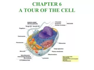
4.4 Eukaryotic cells are partitioned into functional compartments • All other forms of life are composed of more complex eukaryotic cells (four Kingdoms) • Distinguished by the presence of a true nucleus • Membranes form the boundaries of many eukaryotic cells • Compartmentalizing the interior of the cell and facilitating a variety of metabolic activities
Smooth endoplasmicreticulum Nucleus Roughendoplasmicreticulum Flagellum Not in mostplant cells Lysosome Ribosomes Centriole Golgiapparatus Peroxisome Microtubule Plasma membrane Intermediatefilament Cytoskeleton Mitochondrion Microfilament • A typical animal cell • Contains a variety of membranous organelles • May have flagella, but lack a cell wall (what else?) The origin of mitochondria Figure 4.4A
Roughendoplasmicreticulum Nucleus Ribosomes Smoothendoplasmicreticulum Golgiapparatus Microtubule Centralvacuole Intermediatefilament Cytoskeleton Not inanimalcells Microfilament Chloroplast Cell wall Mitochondrion Peroxisome Plasma membrane • A typical plant cell has some structures that an animal cell lacks • Such as chloroplasts and a rigid cell wall (cellulose) • Uusually have a central vacuole and chloroplasts, lack centrioles, and usually lack lysosomes and flagella. The origin of chloroplasts Figure 4.4B
Name: Job: Your Name: __________________ Name: Job: Name: Job: Name: Job: Name: Job: These three are not found in animal cells Collectively, they are called cytoskeleton. Name these three structures and briefly describe their general functions. 1. 2. 3. General Function: Name: Job: Peroxisome Name: Job: Name: Job:
Nucleus Chromatin Two membranesof nuclearenvelope Nucleolus Pore Roughendoplasmicreticulum Ribosomes ORGANELLES OF THE ENDOMEMBRANE SYSTEM 4.5 The nucleus is the cell’s genetic control center • The largest organelle is usually the nucleus (the cellular control center) containing the cell’s DNA, which directs cellular activities • Which is separated from the cytoplasm by the nuclear envelope Figure 4.5
4.6 Overview: Many cell organelles are connected through the endomembrane system • An extensive system of membranous organelles, referred to as the endomembrane system, work together in the synthesis, storage, and export of molecules. • Each of these organelles is bounded by a single membrane. Some are in the form of flattened sacs; some are rounded sacs; and some are tube-shaped. • The major function of the endomembrane system is to divide the cell into separate compartments like an automobile assembly plant
Transport vesiclebuds off 4 Ribosome Secretory(glyco-) proteininside trans-port vesicle 3 Sugar chain 1 2 Glycoprotein Polypeptide Rough ER • 4.8 Rough endoplasmic reticulum makes membrane and proteins • Rough endoplasmic reticulum (rough ER) is composed of flattened sacs that often extend throughout the entire cytoplasm. • The rough ER has three functions: synthesis, modification, and packaging of proteins. • Ribosomes on the surface of rough ER make proteins, some of which are incorporated into the membrane. Other proteins are packaged in membranous sacs that bud off the rough ER and are transported to the Golgi apparatus Figure 4.8
Golgi apparatus “Receiving” side ofGolgi apparatus Golgiapparatus Transportvesiclefrom ER TEM 130,000 New vesicleforming Transportvesicle fromthe Golgi “Shipping” sideof Golgi apparatus • 4.9 The Golgi apparatusfinishes, sorts, and ships cell products • Stacks of membranous sacs receive and modify ER products • Transport vesicles from the ER fuse on one end of a Golgi apparatus to form flattened sacs (Figure 4.9). • These sacs move through the stack like a pile of pancakes added at one end and eaten from the other. Molecular processing occurs in the sacs as they move through the Golgi. • At the far end, modified molecules are released in transport vesicles. Figure 4.9
Smooth ER Rough ER Nuclearenvelope Ribosomes Rough ER Smooth ER TEM 45,000 Figure 4.7 • 4.7 Smooth endoplasmic reticulum has a variety of functions • Smooth endoplasmic reticulum, or smooth ER • Lacks surface ribosomes • Synthesizes lipids • Processes toxins and drugs in liver cells • Stores and releases calcium ions in muscle cells
Rough ER 1 Transport vesicle(containing inactivehydrolytic enzymes) Golgiapparatus Plasmamembrane Lysosomeengulfingdamagedorganelle 2 Engulfmentof particle Lysosomes 3 5 4 Foodvacuole Digestion • 4.10 Lysosomes are digestive compartments within a cell • Lysosomes are sacs of enzymes • Lysosome vesicles contain hydrolytic enzymes that break down the contents of other vesicles, damaged organelles, or bacteria with which they fuse. • Lysosomes are the recycling center for the cell. “Food” Figure 4.10A
Lysosome containingtwo damaged organelles Lysosome Mitochondrion fragment TEM 42,500 Peroxisome fragment Nucleus TEM 8,500 • Lysosomes in white blood cells • Destroy bacteria that have been ingested • Lysosomes also recycle damaged organelles Figure 4.10B Figure 4.10C
Nucleus Chloroplast Centralvacuole Colorized TEM 8,700 • 4.12 Vacuoles function in the general maintenance of the cell • Plant cells contain a large central vacuole that function in storage, play roles in plant cell growth, maintain turgor pressure, and may function as large lysosomes. The vacuoles may also contain pigments or poisons. • Contractile vacuoles in cells of freshwater protists (both protozoa and algae) function in water balance. Figure 4.12A
Transport vesicle fromGolgi to plasma membrane Transport vesiclefrom ER to Golgi Rough ER Plasmamembrane Nucleus Vacuole Lysosome Nuclear envelope Smooth ER Golgi apparatus • 4.13 A review of the endomembrane system • The various organelles of the endomembrane system • Are interconnected structurally and functionally • Vesicles can fuse with the plasma membrane and deliver the content to the extracellular environment without the content actually crossing the plasma membrane. Figure 4.13
Chloroplast Stroma Inner and outermembranes TEM 9,750 Granum Intermembranespace Figure 4.14 ENERGY-CONVERTING ORGANELLES • 4.14 Chloroplastsconver t solar energy to chemical energy • Chloroplasts, found in plants and some protists • Chloroplasts are double-membrane-bounded. • Convert solar energy to chemical energy in sugars, the site of photosynthesis • The capturing of light and electron energizing occur on the granum (plural, grana), and chemical reactions that form food-storage molecules occur in the stroma.
Mitochondrion Outermembrane Intermembranespace Innermembrane TEM 44,880 Cristae Matrix • 4.15 Mitochondria harvest chemical energy from food • Mitochondria carry out cellular respiration, the conversion of glucose to ATP. • found in all cells of eukaryotes, except a few anaerobic protozoans. • are double-membrane-bounded organelles with two membrane spaces, the intermembrane space and the mitochondrial matrix. • the ATP-generating electron transport system is embedded in the inner membrane (cristae), and chemical reactions occur in compartments between membranes. Figure 4.15
Tubulin subunit Actin subunit Fibrous subunits 25 nm 10 nm 7 nm Microtubule Microfilament Intermediate filament THE CYTOSKELETON AND RELATED STRUCTURES • Microfilaments of actin • Enable cells to change shape and move • Intermediate filaments • Reinforce the cell and anchor cer tain organelles • Microtubulesgive the cell rigidity • And provide anchors for organelles and act as tracks for organelle movement
Colorized SEM 4,100 LM 600 Figure 4.17A Figure 4.17B • 4.17 Eukaryotic Cilia and flagella move when microtubules bend • Are locomotor appendages that protrude from minute, tubular extensions of the plasma membrane that surround a complex arrangement of microtubules of certain cells • Are similar in structure. Cilia are short and numerous, while flagella are long and fewer Trachea, oviduct
CELL SURFACES AND JUNCTIONS • 4.19 Cell surfaces protect, support, and join cells • Cells interact with their environments and each other via their surfaces. • Prokaryotic cells and eukaryotic cells of many protists function independently of one another and relate directly to the outside environment. • Animal cells are embedded in an extracellular matrix • Which binds cells together in tissues
Tight junctions Anchoring junction Gap junctions Extracellular matrix Space between cells Plasma membranes of adjacent cells • Tight junctions can bind cells together into leakproof sheets • Anchoring junctions link animal cells into strong tissues • Gap junctions allow substances to flow from cell to cell Figure 4.18B
Walls of two adjacent plant cells Vacuole Plasmodesmata Layers of one plant cell wall Cytoplasm Plasma membrane • Plant cells • Are supported by rigid cell walls made largely of cellulose • Connect by plasmodesmata, which are connecting channels through the cell walls connecting the cytoplasm of adjacent plant cells Figure 4.18A
FUNCTIONAL CATEGORIES OF ORGANELLES • 4.19 Eukaryotic organelles comprise four functional categories • Eukaryotic organelles fall into four functional groups • Manufacture: synthesis of macromolecules and transport within the cell. • Breakdown: elimination and recycling of cellular materials. • Energy processing: conversion of energy from one form to another. • Support, movement, and communication: maintenance of cell shape, anchorage and movement of organelles, and relationships with extracellular environments • All four categories work together as an integrated team, producing the emergent properties at the cellular level.
Eukaryotic organelles and their functions Table 4.19