Download
1 / 12
190 likes | 552 Views
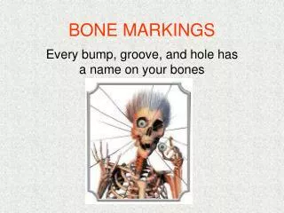
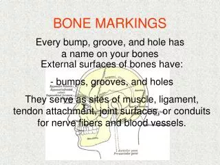
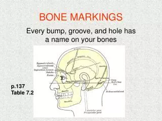
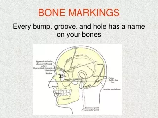
Bone Markings. -Bone markings can be classified as either a depression, a projection that helps forms joints (articulation), or projections that are sites of muscle and ligament attachment. *see page 115 Table 5.1 in text book for further reference.

E N D
Bone Markings -Bone markings can be classified as either a depression, a projection that helps forms joints (articulation), or projections that are sites of muscle and ligament attachment. *see page 115 Table 5.1 in text book for further reference.
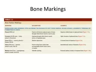
Projections that are sites of muscle and ligament attachment Tuberosity Crest Narrow ridge of bone usually prominent. Palpable Ex. Iliac Crest • Large rounded projection, may be roughened. • Palpable • Ex. Tibial Tuberosity
Trochanter Line Narrow ridge of bone, less prominent than a crest. Not Palpable Intertrochanteric Line • Very large, blunt irregular shaped process. • Palpable • Ex. Greater and lesser Trochanter of the Femur
Tubercle Epicondyle Raised area on or above a condyle. Is a site of ligament attachment Palpable Ex. Medial and Lateral epicondyle of the Humerus • Small rounded projection or process • Palpable • Ex. Greater and lesser tubercle of the Humerus
Spine Ramus Arm-like bar of bone Palpable Ex. Mandibular Ramus • Sharp, slender often pointed projection. • Palpable • Ex. Scapular Spine
Projections that help to form Joints (articulation) Head Facet Smooth nearly flat articular surface. Not Palpable Ex. Rib Facet ( connects to the vertebrae) • Bony expansion carried on a narrow neck. • Not palpable in the Femur or Humerus, but is palpable in the Radius • Ex. Femoral Head
Condyle • Rounded articular projection • Palpable • Ex. Femoral condyle
Depressions (allow passage of blood vessels and nerves) Meatus Sinus Cavity within a bone filled with air and lined with mucous membrane. Not Palpable Ex. Sinus in Cranium • Canal-like passage way • Palpable • Ex. External Auditory Meatus
Fossa Groove Slit-like furrow Not Palpable Ex. Bicipital Groove in Humerus • Shallow basin-like depression in bone, often serving as an articular surface. • Not Palpable • Ex. Glenoid Fossa in Scapula
Fissure Foramen Round or oval opening through bone. Not Palpable Ex. Vertebral Foramen. • Narrow slit-like opening • Allows for blood vessel and nerve passage. • Not Palpable • Ex. Located in Skull
Picture Sources • http://redsports.sg/wp-content/uploads/2008/06/tibial-tuberosity.jpg • http://stemcelldoc.files.wordpress.com/2009/02/iliac_crest_model.jpg • http://www.pediatric-orthopedics.com/Topics/Bones/Femur/Upper_Posterior_Lab.jpg • http://img.tfd.com/vet/thumbs/gr208.jpghttp://www.courses.vcu.edu/DANC291-003/scapula_spine.jpg • http://www.courses.vcu.edu/DANC291-003/scapula_spine.jpg • http://upload.wikimedia.org/wikipedia/commons/7/79/Femur_head.png • http://1.bp.blogspot.com/_15-2o9FAeCE/SATS864PRJI/AAAAAAAAAN0/fcFKrwwO52A/s400/rib1.jpg • http://www.health-res.com/EX/07-28-00/knee_OCD_anatomy01.jpg • http://upload.wikimedia.org/wikipedia/commons/5/52/Human_skull_lateral_view.jpg • http://www.health.com/health/static/hw/media/medical/hw/n1808.jpg • http://www.chionline.com/anatomy/anat37.gif • http://mial.fas.sfu.ca/Files/BGHumerus.jpg • http://en.wikivisual.com/images/4/4e/Gray_190_-_The_skull_from_the_front.png http://www.apparelyzed.com/_images/content/spine/vertebrae-spine.jpg
Bibliography • Marieb, E. N. (2000). Overview of the Skeleton. Essentials of Human Anatomy and Physiology (pp. 47). Reading: An Imprint of Addison Wesley Longman Inc. .