Download
1 / 43
460 likes | 792 Views
Echo Club Invasive hemodynamic evaluation of regurgitation. Mohammed Almansori MBBS, FRCPC Assistant Professor of Medicine & Interventional C ardiologist University of Dammam.

E N D
Echo ClubInvasive hemodynamic evaluation of regurgitation Mohammed Almansori MBBS, FRCPC Assistant Professor of Medicine & Interventional Cardiologist University of Dammam
In every patient in whom it was considered that a decision could be reached by echocardiography alone (>80% of pts) there was 100% agreement from cardiac catheterization. • So, in patients with adequate echo data Cath can be omitted. • If echo indices are conflicting or significant CAD is suspected catheterization should be considered
Indication of invasive hemodynamic evaluation: When pulmonary pressure is disproportionate to the severity of regurgitation assessed noninvasively. When there is a discrepancy between clinical and noninvasive findings.
Reg stroke volume = Angio stroke volume – Forward stroke volume Rough estimation of RF compared to visual interpretation: 1+ Reg is equivalent to RF of 20% 2+ Reg is equivalent to RF of 20-40% 3+ Reg is equivalent to RF of 40-60 4+ Reg is equivalent to RF of > 60%
Prominent V wave Peak > 40 mmHg Delta peak V wave and mean PCWP > 10 mmHg Ratio peak V wave and mean PCWP > 2 It does not correlate with the presence or severity of mitral regurgitation
A small diastolic pressure gradient may be observed across the mitral valve; however, unlike mitral stenosis, the gradient is present during early diastole only. In addition, the slope of the y descent in mitral regurgitation is steep rather than delayed, as seen in mitral stenosis.
The transmission of this pressure wave from the pulmonary veins to the pulmonary artery also explains the occasional phenomenon of a false elevation in the pulmonary artery saturation, in some cases of severe mitral regurgitation.
In patients with only minor mitral regurgitation which is suspected to contribute to their clinical symptoms , the monitoring of invasive hemodynamic parameters (V-wave) during stress is important
A 71-year-old man know to have moderate AR presented with a month of CCS-III angina and shortness of breath. No other medical illnesses. O/E HR 63 BP 164/53 JVP 9 cm H2O Early diastolic murmur of AR + Pansystolic murmur of MR + S3 Occasional crepitation on chest auscultation No LL edema CXR cardiomegaly , No pulmonary edema
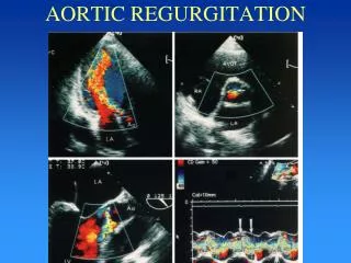
Echo: Normal LV function. (LVIDd 6.3cm - LVIDs 4.0cm) Moderate to severe AR Moderate MR
Coronary angiogram showed mild non-obstructive disease. Aortic root angiogram showed severe aortic regurgitation.
Mean PAP 42 mmHg PCWP 25 mmHg Cold not get LV tracing due to catheter induced VT