Download
1 / 36
360 likes | 635 Views
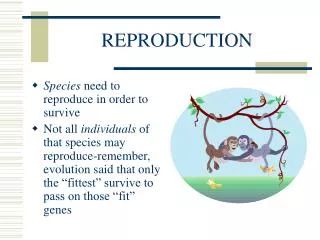
Continuity Through Reproduction. Chapter 6. Types of Reproduction. Sexual – reproduction involving 2 different cells, usually from 2 parents. Asexual – new individuals originate from a single parent. Amoeba. Vegetative – asexual reproduction in plants.

E N D
Continuity Through Reproduction Chapter 6
Types of Reproduction Sexual – reproduction involving 2 different cells, usually from 2 parents.
Asexual – new individuals originate from a single parent. Amoeba
Regeneration – ability of an organism to replace lost body parts. planaria
Reproduction – life continuing from one generation to the next.
Gametes (sex cells) Sperm cell • Produces by males • Consists of a little more than a nucleus, a tail that can move the cell about, and an energy generator in its mitochondria
Egg cell (Ovum) • Produced by females • Maybe large, sometimes thousands of times larger than a sperm cell • It often contains a reserve food supply
Female Organism that produces ova
Male Organism that produces sperm
Fertilization • The joining of an ovum & sperm • Sperm penetrates the ovum’s outer membrane & enters its cytoplasm • Zygote: a fertilized egg
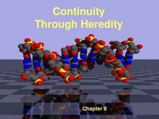
Genome The total genetic content of the chromosomes of the cell. In human cells, the genome consists of 23 pairs of chromosomes.
Homolog Each member of a chromosome pair A pr. Of chromosomes consists of 2 homolog that are similar in appearance.
Haploid – one set of chromosomes the N number of chromosomes the gametes are haploid cells
Diploid – the total # of chromosomes in the zygote and all body cells the 2N number
Meiosis • Special cell division process where the # of chromosomes is reduced from diploid to haploid • Stages are similar to Mitosis • Occurs in special reproductive organs • Testes – males • Ovaries – females • there are 2 nuclear divisions, Meiosis I & Meiosis II
Meiosis I – Reduction Division Prophase I • Long, thin chromosomes shorten & thicken • Each homolog finds it’s partner & they pr. w/one another along their entire lengths • Homologs lie very close & are often twisted around one another • Chromatids actually can break at various places & join with broken chromatids from the other homologs. Known as crossing over and results in recombination of genes. • Toward the end, the chromosome prs. Move to the equatorial plate
Metaphase I • Chromosome pairs arrive in the center of the cell
Anaphase I • The homologs of each pair separate & begin to move toward opposite poles of the spindle • Each chromosome still consists of 2 chromatids, attached at the centromere
Telophase I • Cytokinesis occurs & 2 cells are formed • Each new cell has only half as many chromosomes as in a body cell. Each new cell has only half the parent cell’s total genetic info.
Meiosis II • Usually begins almost immediately • There is no new replication of DNA in the period between Meiosis I & Meiosis II
Prophase II • Remaining chromosomes move toward the equatorial plate on a new spindle • The 2 chromatids of each chromosome are about to separate at the centromere
Metaphase II • Chromosomes align themselves on the new equatorial plate
Anaphase II • The centromeres divide & the 2 chromatids of each chromosome separate & move towards opposite poles • Each chromatid is now an independent chromosomes from this time forth
Telophase II • Chromosomes gather at the poles & are enclosed by a new nuclear envelope • Cytoplasm divides again • Cells undergo further cytoplasmic changes, such as developing a tail • 4 mature sperm cells are formed only 1 mature egg cell is formed Polar bodies: tiny cells w/very little cytoplasm produced in meiosis which are expelled.
Males: Testes develop in the abdominal cavity of an embryo, just like the ovaries do. Before birth, the 2 testes move downward & then to the outside of the abdominal cavity. The testes are housed in a pouch called the scrotum. The scrotum is located just below the external male organ, the penis – used for both reproduction & for discharging urine from the kidneys. Each testes is made up of thousands of tiny tubules. Meiosis occurs in these tubules.
Sperm moves out of the canals into a special collecting duct near the testes known as the epididymis. They remain there until they are released in sexual activity or until they are reabsorbed by the body. A male may release from 200 million to 500 million sperm cells at one time. Gametes are produced at puberty.
Females: 2 ovaries are located low n the abdominal cavity, one on each side. Each ovary is located close to a short tube, the oviduct. Oviducts serve as passageways and ova travel through them on their way to the uterus, a muscular organ that protects & nourishes a new individual while it develops. Each potential ovum is enclosed in a follicle, a small oblong sac of cells on the surface of the ovary. During the 10 days before an ovum is released, estrogen from the follicle stimulates the inner layer of the uterus.
Estrogen: hormone which promotes the development of the inner tissues of the uterus. After ovum is released, the follicle cells fill the follicle cavity & appear yellow in color, known as the Corpus Luteum. Progesterone: produced my the corpus luteum. It helps maintain the thick layer of the uterus for 2 more weeks, enough time for the embryo to attach itself to the uterine wall. If the egg is not fertilized, the thick layer disintegrates. The corpus luteum stops producing progesterone. Inner layer of the uterus breaks away & passes through the vagina, a muscular passageway that connects the uterus to the outside.
Menstruation The loss of blood & tissue from the uterus. The flow lasts for 4 to 5 days. Menstrual Cycle: a regular monthly cycle initiated by the brain & ovarian hormones.
Hormones Chemical messengers that influence other organs. They travel in the blood to all parts of the body. GH - pituitary Thyroxin - thyroid Testosterone - testes Adrenalin - adrenal Insulin - pancreas
Sperm cells begin to move from the epididymus inot a special duct, vas deferens. The vas deferens enters the duct of the penis. The male reproductive system expels 3 – 4 mL. of semen – whitish fluid containing secretions of the male glands (prostate glands, seminal vesicles, and Cowper’s gland.) Sperm cells, swimming in all directions, enter the uterus & two oviducts. After ovum is released, it moves into the nearest oviduct. It will live 3 – 5 days.
One sperm cell penetrates the outer membranes of the egg cell. Once a sperm cell enters the ovum, the membranes react & become a barrier to other sperm cells. Development of new individual begins in the oviduct.