Download
1 / 41
410 likes | 444 Views
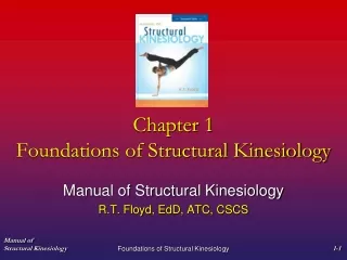
Foundations of Structural Kinesiology. Chapter 1. And you thought this class was stressful. Skeletal System. Skeletal System Divisions (206 bones). Axial: (80 bones) Skull Spinal column Sternum Ribs Apendicular: (126) Shoulder Girdle Upper Extremity Pelvic Girdle Lower Extremity.

E N D
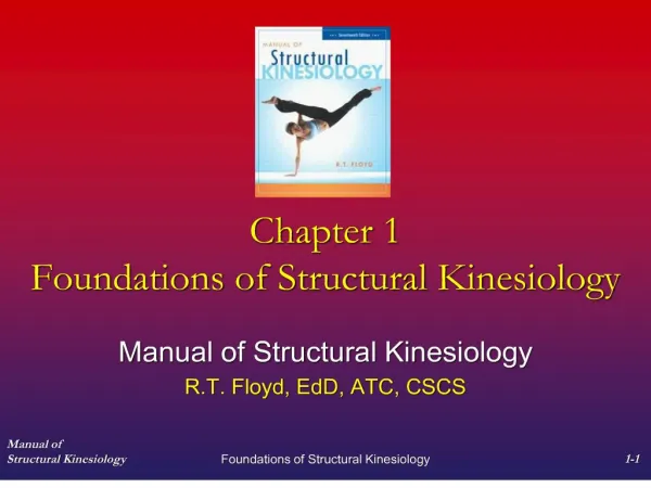
Foundations of Structural Kinesiology Chapter 1 And you thought this class was stressful
Skeletal SystemDivisions (206 bones) • Axial: (80 bones) • Skull • Spinal column • Sternum • Ribs • Apendicular: (126) • Shoulder Girdle • Upper Extremity • Pelvic Girdle • Lower Extremity
Skeletal System • Functions • Support • Protection • Movement • Storage • Hemopoiesis
Bone Properties • Bone size (mass) and shape • formation according to the stresses (direction and magnitude of force) habitually applied to them • Wolff’s Law Athlete Scoliosis Torsion
Skull Vomer Parietal Nasal Conchae Palatine Frontal Sphenoid Ethmoid Occipital Lacrimal Nasal External Occipital Protuberance Maxilla Temporal Vomer Zygomatic Sphenoid Mastoid Process Mandible Superior Nuchal Line(3rd picture #2) Label
Vertebra Intervertebral Foramen Spinous Process Articular Processes Lamina Transverse Processes Pedicle Vertebral Foramen Body Label
Regional Structures Cervical (7) Bifed Spinous Process Transverse Foramen Atlas Axis (Odontoid Process) Thoracic (12) . . Lumbar (5) . Median Sacral Crest Sacral Canal Sacral Foramen Sacral Hiatus Superior Articular Facet Superior Sacral Notch Coccyx (4) Vertebral Column Bifed Spinous Process • Superior & Inferior • Costal Facets • Transverse Costal Facets Sacral (5)
Sternum Jugular Notch Clavicular Notch Manubrium Sternal Angle Costal Notches Body Transverse Ridges Xyphoid Process LABEL
Rib Cage Superior Border Tubercle Neck Costal Angle Head Inferior Border True (7) Costal Groove Shaft False (3) Floating (2) Label Label
Clavicle Deltoid Tubercle Acromial End Superior surface Anterior Sternal End Anterior Trapezoid Line Inferior surface Right Clavicle Subclavian Groove Conoid Tubercle Label
Scapula Supra Scapular Notch Superior Angle Coracoid Process Supraspinous Fossa Spine Supra Glenoid Tubercle Superior Border Acromion Process Glenoid Fossa Infra Glenoid Tubercle Medial (vertebral) Border Infraspinous Fossa Inferior Angle Lateral (axillary) Border Label
Humerus Greater Tubercle Head Anatomical Neck Lesser Tubercle Surgical Neck Intertubercular Groove (bicipital) Deltoid Tuberosity Radial Groove Olecranon Fossa Supracondylar Ridge Radial Fossa Lateral Epicondyle Capitulum Anterior Posterior Coronoid Fossa Trochlea Medial Epicondyle Label
Radius and Ulna Olecranon Process Semilunar Notch Radial Notch Head Coronoid Process Neck Radial Tuberosity Ulnar Tuberosity Supinator Crest Interosseous Crest Supinator Fossa Ulnar Notch Styloid Process Head Styloid Process Radius Label Ulna Label
Hand Distal Middle Phalanges Proximal Head Shaft Metacarpals Hamate (Hook of Hamate) Base Trapezoid Pisiform Carpals Trapezum Triquetrum Label Scaphoid Capitate Lunate
Posterior Gluteal Line Inferior Gluteal Line Pelvic Girdle External Surface Anterior Gluteal Line Iliopectineal Eminence Iliac Crest Iliac Fossa Posterior Superior Iliac Spine Ischial Tuberosity Superior Rami Pubic Crest Posterior Inferior Iliac Spine Spine of the Ischium Superior Rami Anterior Inferior Iliac Spine Auricular Surface Anterior Superior Iliac Spine Lesser Sciatic Notch Inferior Rami Inferior Rami Greater Sciatic Notch Sacral Articulation Pubic Symphysis Ilium Label Ischium Label Pubis Label
Femur Greater Trochanter Quadrate Tubercle Fovea Capitus Head Intertrochanteric Crest Neck Intertrochanteric Line Gluteal Tuberosity Lesser Trochanter Linea Aspera Adductor Tubercle Popliteal Surface Intercondyloid Fossa Patellar Surface Lateral condyle Medial condyle Right Femur (Anterior) Right Femur (Posterior) Label
Patella Proximal Border Base Lateral Border Medial Border Apex Right Patella Label
Tibia & Fibula Intercondyloid Eminence Styloid Process Superior Fibular Articulation Lateral Condyle Medial Condyle Tibial Tuberosity Head Soleal Line Interosseous Border Anterior Border Interosseus Border Anterior Border Inferior Fibular Articulation Medial Malleolus Lateral Malleolus Talus Articulation Talus Articulation Fibula Label Tibia Label
Bones of the Foot Distal Middle Proximal Phalanges Head Talus Shaft Navicular Base Metatarsals 1st Cuniform 1st Cuniform Cuboid 2nd Cuniform Calcaneus 3rd Cuniform Tarsals Navicular Talus Label Calcaneus
Soft/Connective Tissues 1. Ligament 2. Tendon 3. Retinaculum 4. Fascia 5. Aponeurosis 6. Cartilage 7. Meniscus 8. Labrum - A dense connective tissue that encloses, separates, and binds muscles – supports, protects, and gives shape. Also gives general support to an area - Crescent plates of fibrocartilage that deepen an articular surface and act like shock absorbers - A dense connective tissue sheath that binds or holds tendons in place - A tendinous connective tissue formed like a sheath that anchors one muscle to another - Deepens a socket (articular capsule blends into labrum and surrounds anatomical neck) - A band of dense connective tissue that connects a muscle to a bone or other structure - A firm, smooth, resilient, nonvascular connective tissue - A thick fibrous band of connective tissue that connects bone to bone
Anatomical Position & Fundamental Position Anatomical Position Fundamental Position
Reference Segment Reference Side Reference Side & Segment
Anatomical Directional Terminology • Anterior • (Anteroinferior, Anerosuperior, • Anterolateral, Anteromedial, • Anteroposterior) • Posterior • (Posteroinferior, • Posterosuperior, Posterolateral, • Posteromedial, Posteroanterior) • Superior • (Superolateral, Superomedial) • Inferior • (Inferolateral, Inferomedial) • Medial • Lateral • Ipsilateral • Contralateral • Unilateral • Bilateral • Caudal • Cephalic • Dorsal • Ventral • Proximal • Distal • Ventral • Proximal • Distal • Superficial • Deep • Palmer • Volar • Prone • Supine • Dorsum
Articular SystemThe union/articulation of two or more bones • Types of Joints- Classifications of articulations grouped according to structure or function • Structural: Fibrous, Cartilaginous, Synovial • Functional: • Synarthrodial (immoveable) • Suture • Gomphosis
Articular System • Amphiarthrodial (slightly moveable) • Syndesmosis- held together by strong ligaments • Symphasis- separated by fibrocartilage pad • Synchrondrosis- separated by hyaline cartilage • Diarthrodial (Freely moveable) • joint cavity, joint capsule, synovial membrane, synovial fluid, articular cartilage
Ginglymus (hinge) -Uniaxial Interphalangeal Metacarpophalangeal of thumb Humeroulnar Tibiotarsal Femorotibial (dual) Seller (Saddle) -Multiaxial Carpometacarpal of thumb Enarthrodial (Ball & Socket) -Multiaxial Acetabulofemoral Glenohumeral Diarthrodial Joints Arthrodial (Gliding) -Multiaxial • Carpal articulations • Tarsal articulations • Acromioclavicular • Sternoclavicular • Patellofemoral • Radiohumeral (pivot?) • Vertebral art. Process • Carpometacarpal • Tarsometatarsal • Sup. & Inf. Tibiofibular • Sternocostal • Costovertebral • Costotransverse • Intermetatarsal • Intermetacarpal Trochoidial (Pivot) -Uniaxial • Radioulnar (prox & dist) • Atlantoaxial Condylodial (Convex-Concave) -Biaxial • Metacarpophalangeal • Radiocarpal • Atlantooccipital • Femorotibial (dual) • Metatarsophalangeal
Movement in Joints • Motion that takes place by the bones moving through a plane of motion about an axis is referred to as physiological movement or osteokinematic motion. • Movement Terminology are the terms used to describe the actual change in position of the bones relative to each other. • The specific amount of movement in a joint can be measured using an instrument called a goniometer.
General Movements Abduction Diagonal Abduction Adduction Diagonal Adduction Flexion Internal Rotation Extension External Rotation Circumduction Hyperextension Anatomical Movement Terms Specific to the Ankle and Foot Eversion Dorsiflexion Inversion Plantarflexion
Anatomical Movement Terms Specific to Shoulder Girdle & Shoulder Joints Depression Elevation Protraction (Scapular Abduction) Retraction (Scapular adduction) Upward Rotation Downward Rotation Horizontal Abduction (shoulder joint) Horizontal Adduction (shoulder joint) Specific to the Radioulnar Joints Pronation Supination
Anatomical Movement Terms Specific to the Spine Lateral Flexion Reduction Rotation Specific to the Wrist and Hand Dorsal Flexion (ext.) Long Abduction Palmar Flexion (flex.) Short Abduction Radial Deviation Thumb Oppostion Ulnar Deviation Thumb Reposition Specific to the Mandible Protrusion Retrusion
Arthrokinematic Motion • In order for physiological movements to occur there must be movement between the actual articular surfaces of the joint. This is known as arthrokinematic motion. There are three specific types: • Roll (rock)- a series of points on one articulating surface contacts with a series of points on another articular surface • Glide (slide, translation)- a specific point on one articulating surface comes in contact with a series of points on another surface) • Spin– A single point on one articular surface rotates about a single point on another articular surface.