Download
1 / 71
710 likes | 713 Views
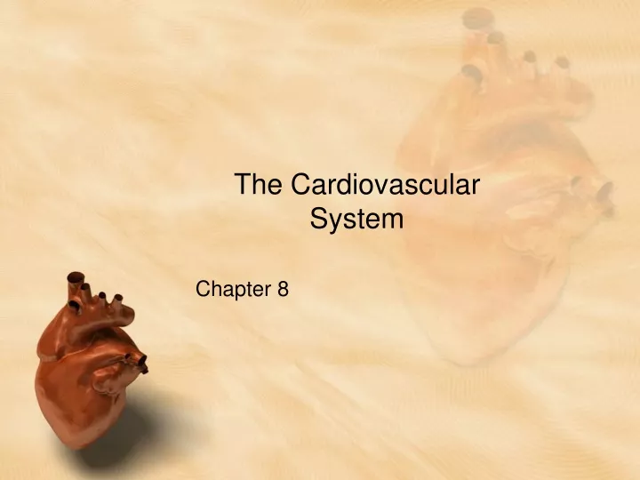
The Cardiovascular System. Chapter 8. http://www.youtube.com/watch?v=upctPUa6RhA. Pump it Up!!!. The heart is a pump for delivery of: Oxygen Nutrients Hormones Antibodies WBC’s Also removes wastes and antioxidants.

E N D
The Cardiovascular System Chapter 8
Pump it Up!!! • The heart is a pump for delivery of: • Oxygen • Nutrients • Hormones • Antibodies • WBC’s • Also removes wastes and antioxidants. • All of these materials are propelled by the heart through a closed system of tubes to the tissues of the body. • What are these tubes called?
Where is the heart located?? • Centrally located in the chest. • Surrounded by lungs • Protected by ribs • Seems to be slightly shifted to left side of the chest • Heart lies in the mediastinum- space between the pleural cavities that contain the lungs. Also called the interpleural space. • Trachea, esophagus, and other vascular structures are also contained in the mediastinum.
Basic Heart Terminology. • Apex would be considered at the bottom of the heart near the ventricles. • Base is at the top of the heart, where major blood vessels enter and exit.
External Structures of the heart • Auricles- largest and most visible parts of the atria. • Ventricles are separated by interventricular sulci. • Atria do not have as thick of walls as the ventricles do. Why?? • Remember that there are vessels that supply the heart with blood itself as well. This is called coronary circulation • Highest pressure is found in aorta. Why??? • Brachiocephalic trunk and left subclavian artery branch off aorta just after aortic valve.
Composition of the Heart Wall • Primarily a muscle. • Outer layer is called the pericardium. • Consists of two layers with fluid filled cavity between. • 1. Outer fibrous pericardium • Made of tough, fibrous connective tissue that protects the heart and loosely attaches to the diaphragm. • 2. Inner serous pericardium • Actually made up of two layers • Inner visceral layer called the epicardium. • Outer parietal layer
Composition of Heart Walls continued… • Inside the sac formed by the pericardium is the myocardium- the thickest layer of the heart tissue. • Between the myocardium and the heart chambers is a thin membranous lining called the endocardium.
Internal Structures of the Heart • The Valves of the heart • Right Atrioventricular Valve (also called right AV valve or tricuspid valve). • Left atrioventricular Valve (also called the left AV valve or mitral valve or bicuspid valve). • Pulmonary valve (also called pulmonic valve is a semilunar valve). • Aortic valve (is a semilunar valve).
Valve Composition • Have 2 or 3 leaflets (flaps) that originate from the annulus of the valve which is a fibrous ring. • These are the outer edges of the flaps • Inner edges of flaps are attached to papillary muscles by chordae tendinae. • In right ventricle, there is a band of tissue that originates at the interventricular septum but does not attach to the flaps of the tricuspid valve; it is called the Moderator band and connects to the outside wall of the right ventricle.
So how does this all work??Atrial Contraction/Ventricular Relaxation
Blood Flow through the heart • Let’s Review • What do veins do? • What do arteries?
Blood Flow through the heart continued…. • Blood only flows in one direction in a healthy heart. • Basic function is to receive deoxygenated blood from the tissues of the body, pumps it through the lungs and then back out through the body system.
Blood Flow Steps • 1. Caudal or cranial vena cava • 2. Right atrium • 3. Right Atrioventricular (AV) valve (tricuspid valve). • 4. Right ventricle • 5. Pulmonary valve • 6. Pulmonary arteries • 7. Lungs • Exchange takes place at alveoli/capillaries • 8. Pulmonary veins • 9. Left atrium • 10. Left Atrioventricular (AV) valve (mitral valve). • 11. Left Ventricle • 12. Aortic Valve • 13. Aorta • 14. Systemic capillaries • 15. Tissue • 16. Back to caudal or cranial vena cava
Phases of blood flow through the heart. • Systole- mitral and tricuspid valves close and ventricles pumps blood out pulmonic valve and aortic valves. • Diastole- Ventricles refill with blood with tricuspid and mitral valves open and pulmonic and aortic valves closed.
http://www.sumanasinc.com/webcontent/animations/content/human_heart.htmlhttp://www.sumanasinc.com/webcontent/animations/content/human_heart.html
Heart Sounds…LUB DUB • “Lub” sound of heart is also called S1. • Closing of the AV valves • “Dub” sound is also called S2. • Closing of the semilunar valves. Where do we listen for these sounds??
The Cardiac Cycle • What causes the heart to actually pump? • Electrical impulse for heartbeat comes from the sinoatrial node (SA node) located in the right atrium and known as the pacemaker for the heart. • SA node is a specialized area of cardiac muscle cells that can generate automatically the impulses that trigger the repeated beating of the heart.
How is electrical impulse generated? • Remember Depolarization/Repolarization? • Polarization: Cations (substances with a positive charge) are pumped out of the cell. This results in in outside of cell having a more positive charge than inside cell. • Depolorization: Gates open to allow cations to flow back into cell to equalize charge. This generates an electrical current which causes the heart to contract. • Depolarization= Systole • Repolarization= Diastole
Electrical Activity Continued • Electrical current is generated in SA node and travels one of two paths from base of heart to apex of heart. • Speedy route-through cardiac muscle to AV node and Purkinje fibers • Scenic route- Through cardiac muscle fibers alone. • Cardiac muscle can generate electrical impulse from one muscle cell to another, so electrical impulses spread like a ripple through the heart.
Electrical Activity Continued • Electrical Impulse is generated in SA node and then spreads to atria. • Atria contract pushing blood through AV valves to ventricles. • Impulse travels to AV node where it is delayed until atrial systole is complete. • After AV node, electrical impulse travels through specialized fibers in ventricles known as Bundle of His and the Purkinje Fibers. • Purkinje fibers carry impulse into ventricular myocardium.
1 Sinoatrial node (Pacemaker)2 Atrioventricular node3 Atrioventricular Bundle (Bundle of His)4 Left & Right Bundle branches5 Purkinje Fibers
http://www.nhlbi.nih.gov/health/dci/Diseases/hhw/hhw_electrical.htmlhttp://www.nhlbi.nih.gov/health/dci/Diseases/hhw/hhw_electrical.html
The Electrocardiogram • An electrocardiograph or EKG (most correctly termed ECG) is used to detect the electrical activity associated with the heart cycle. • The ECG is useful in detecting abnormalites of the heart based on the graphical appearance.
Interpreting an ECG • P-wave- when the atria contract or depolarize. • QRS complex- when the ventricles depolarize. • T- wave- The repolarization of the ventricles.
www.nhlbi.nih.gov/health/dci/Diseases/hhw/hhw_electrical.htmlwww.nhlbi.nih.gov/health/dci/Diseases/hhw/hhw_electrical.html
Blood Circulation in the Fetus • Major difference in blood flow is that newborn receives oxygen through its own lungs while fetus receives oxygen from blood of mother. • Blood therefore bypasses the lungs during the cardiac cycle in a fetus. • Fetus receives oxygen through the placenta. • Blood from umbilical vein flows through liver (some bypasses liver via ductusvenousus), into caudal vena cava, then into right atrium.
Fetal Circulation Continued • Two forms of bypass in the fetus. • Foramen ovale- between right and left atria. • Ductusarteriosis-if blood flows into right ventricle, then will go from pulmonary artery to aorta. • Deoxygenated blood is sent back to placenta via umbilical arteries to become oxygenated from mother. • At birth, lungs inflate and the newborn will oxygenate its own blood. Normally all bypasses will close at this point.
Heart Rate and Cardiac Output • Cardiac output- the amount of blood that leaves the heart. • Must be sufficient for life sustaining activities. • Is determined by 2 factors: • Stroke volume • Amount of blood ejected with each cardiac contraction • Heart rate • How often the heart contracts. • Is expressed: • Cardiac Output (CO)=Stroke Volume (SV) x Heart Rate (HR)
How to calculate Cardiac output. • If a dog ejects 4 mls of blood with each systolic contraction and Heart rate is 120 bpm. • CO= 4x120 • CO=480 mls/min How does this relate to large animals??
Cardiac output continued • Vigorous exercise increases the demand for oxygen in the tissues, so cardiac output must increase to meet that demand. • This process is called increased contractility or positive inotropy. This in turn will increase stroke volume. • So basically during exercise, increased heart rate, increased cardiac output will increase stroke volume.