Download

1 / 25
250 likes | 322 Views
General Structure of the Digestive Tract. common structural characteristics four main layers: the mucosa, submucosa , muscularis , and serosa . The mucosa- mucous membrane. an epithelial lining;

E N D
General Structure of the Digestive Tract • common structural characteristics • four main layers: the mucosa, submucosa, muscularis, and serosa.
The mucosa- mucous membrane. • an epithelial lining; • an underlying lamina propria of loose connective tissue rich in blood vessels, lymphatics, lymphocytes and smooth muscle cells, sometimes also containing glands • thin layer of smooth muscle called the muscularismucosae usually separating mucosa from submucosa.
Main functions of epithelial lining • Selectively permeable barrier between the contents of the tract and the tissues of the body, • Facilitate the transport and digestion of food, • Promote the absorption of the products of this digestion, • Producehormonesthat affect the activity of the digestive system, • Producemucus for lubrication and protection.
The submucosa • denser connective tissue • many blood and lymph vessels • submucosalplexus of autonomic nerves • may also contain glands and lymphoid tissue
The abundant lymphoid nodules in the lamina propria and the submucosal layer protect (in association with the epithelium) from bacterial invasion • entire digestive tract—with the exception of the oral cavity, esophagus, and anal canal—is lined by a simple thin, vulnerable epithelium.
The lamina propria, located just below the epithelium, is a zone rich in macrophages and lymphocytes, some of which actively produce antibodies. • These antibodies are mainly immunoglobulin A (IgA) and are secreted into the intestinal lumen bound to a secretory protein produced by the epithelial cells.
This complex protects against viral and bacterial invasion. • IgA is resistant to proteolytic enzymes and can therefore coexist with the proteases present in the lumen.
The muscularismucosae allows local movements of the mucosa independent of other movements of the digestive tract, increasing contact of the lining with food. • The contractions of the muscularis, generated and coordinated by autonomic nerve plexuses, propel and mix the food in the digestive tract.
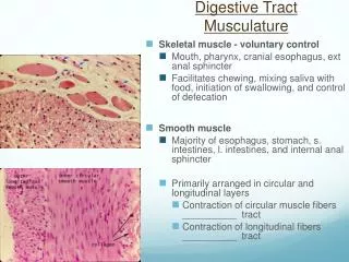
muscularis • thick • smooth muscle cells -spirally oriented and divided into two sublayers. • internal sublayer (closer to the lumen), the orientation is generally circular • external sublayer, it is mostly longitudinal.
In the connective tissue between the muscle sublayers are • blood and lymph vessels • autonomic myenteric nerve plexus. • This and the submucosal plexus together comprise the local enteric nervous system of the digestive tract, containing largely autonomic neurons functioning independently of the central nervous system (CNS).
The serosa • thin layer of loose connective tissue • rich in blood vessels, lymphatics, and adipose tissue • simple squamous covering epithelium (mesothelium)
In the abdominal cavity, the serosa is continuous with • the mesenteries (thin membranes covered by mesothelium on both sides), which support the intestines • peritoneum, a serous membrane that lines the cavity.
In places where the digestive tract is not suspended in a cavity but bound to other structures, such as in the esophagus the serosa is replaced by a thick adventitia, consisting of connective tissue containing vessels and nerves, lacking mesothelium.
Transverse section showing the muscularis halfway along the esophagus reveals a combination of skeletal muscle (right) and smooth muscle fibers (left) in the outer layer, which are cut both longitudinally and transversely here. This transition from muscles under voluntary control to the type controlled autonomically is important in the swallowing mechanism.