Download
1 / 77
780 likes | 1.32k Views
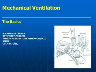
Modes and initiation of ventilation By Elizabeth Kelley Buzbee AAS, RRT-NPS. Highlights of Mechanical ventilation Unit 4. The modes of ventilation:.

E N D
Modes and initiation of ventilation By Elizabeth Kelley Buzbee AAS, RRT-NPS Highlights of Mechanical ventilation Unit 4
The modes of ventilation: • A spontaneous breath is one that the patient triggers and cycles the breath, and he controls the VT . This breath could be assisted by the application of positive pressure. • A mandatory breath is defined as one that is triggered and cycled by the machine. All mandatory breaths are assisted breaths.
The modes of ventilation: full support modes • CMV: continuous mandatory ventilation in which all breaths are mandatory. • VC-CMV volume control also called Assist/Control mode • Set VT, f to get VE; guaranteed VT • Default ventilatory mode for full support with adults • PC-CMV pressure control mode. Patient can trigger breaths just like with A/C • Set PIP, f and TI no guaranteed VT • Default ventilator mode for full support for infants
Indications for PC: the RCP selects pressure ventilation when: • The adult patient who cannot be managed with VC In this case, we keep the PIP less than 30 cmH20. • PC results in better distribution of ventilation in persons with unequal RAW, but consistent compliances. • There is such an airway leak so that the VT are unstable [most common with infants and small children with uncuffed ET or tracheostomy tubes]
Compare PC to VC • In PC, the airway pressures; mPAW and PIP will stay the same, but the VE and VT can vary based on patient’s time constants • In VC the VE and VT are basically stable [patient can increase f so VE could vary] the PIP and the mPAW can be altered by patient time constants
Compare control mode to Assist/control • We control patients by giving them sedation and paralytic agents so that the VE we set on VC-CMV is exactly the same • We can control their PaC02 thus their acid base balance • In A/C, the patient can trigger breaths that will increase the VE, so that the VE based on set VT and f could be lower than the actual measured VE
Controlling the chronic hypercapnic patient • If your patient has a hypoxic drive, administrate enough Fi02 to get his Pa02 between 80-100 mmHg. • This will result in apnea and works as a form of sedation in the first 24 hours. • Must wean the Fi02 to get Pa02 between 55-65 mmHg before weaning
Problems with A/C • Excessively high PAW can cause problems with hemodynamics once patient starts to breath. • Another problem with A/C mode is the risk of auto-PEEP and air trapping.
Inverse Ratio Ventilation [with PC or with VC] • This is a form of full support that uses increased Ti to raise the mPAW when patient’s compliance is so bad that PIP and Pplateau are excessive • In IRV, the expiratory time is so short that the patient never completely exhales. This works like PEEP to recruit alveoli
Raising mPAW with IRV • mPAW = PIP [I] + PEEP [E] • I + E • Because we raise the inspiratory time so much we can decrease the PIP • Because we create auto-PEEP with the short TE, we can decrease the PEEP
Negative pressure ventilation • The negative pressure ventilator is a box in which the patient’s body [or chest wall] is placed. A suction device is attached to the box. • The NPV merely replaces the ventilatory muscles.
Problems with Negative pressure Ventilation: patient must be able to: • protect airway • Handle being supine all the time • hemodynamically stable • be comfortable in one position all the time • handle being disconnected from vacuum for short time spans
More problems with NPV • Patient can get skin lesions from movement of body inside the device • Patient can get cold from ‘wind’ • Best 02 device is nasal cannula because 02 can be sucked into the neck opening
NPV • Classified as controllers, but newer models can be A/C if there is a flow sensor placed on the patient’s nose • Old metal iron lungs have a constant I:E of I:I; newer fiberglass devices can have altered I:E ratios
Setting parameters on NPV: • Change level of the vacuum to increase the VT [he could use a Wright’s spirometer attached to an IPPB mask to measure exhaled VT] • Change the respiratory rate.
CSV • continuous spontaneous ventilation in which all breaths are spontaneous. • patient who can completely control his VE • and only needs a little help such as with increased baseline pressures [CPAP] • or some application of assisted breaths such as pressure support [PS] • or who might require monitoring of VE
Pressure support ventilation • PSV is the most common form of pressure cycled CSV. • Although this does raise the airway pressure so that we have a higher and lower pressure, we call this PS rather than PIP because of the specific characteristics of PS • Flow triggered and flow cycled • Patient controls his VT, f and inspiratory time
Indications for PS: • When used with SIMV to reduce the WOB by increasing the spontaneous VT. We generally select the PS that will deliver a reasonable VT [watch the spontaneous RR] • Can be used alone during weaning. Once a patient is on a PS of 5-10 cmH20, he is considered at a level that only compensates for RAW of the tubing, so is considered consistent with spontaneous breathing.
PSV flow patterns • The flow pattern is descending till it reaches 5 LPM [or 25% of the peak flow] in which the flow stops abruptly. • The flow slows down as the device attempts to keep the PS at the preset pressure.
VT on PS • There is no guaranteed VT, nor VE, but we can increase the VT by increasing the PS pressure • We need to set VE & high f alarms closely to warn us of problems • The patient sends more air to Zone III because he is using his diaphragm more with PS
To choose the correct level of PSV there are three methods: • get an appropriate VT [10-15 ml/kg] and titrate the PS level to achieve this VT • increase the PS level till the respiratory rate is normalized [25 bpm or less] • increase the PSV until you decrease the work of breathing through the ET tube • To select the appropriate level of PSV to overcome the RAW use this formula PSV= (PIP - Pplateau) x spont insp. Flow rate [l/sec] Ventilator flow rate [l/sec]
PSmax • or ‘straight pressure support’ or ‘stand alone PS’ [ PS without SIMV.] In this case, the PS is not used as a weaning modality but for initial of mechanical ventilation. • We generally select a PS level that will deliver 10-12 ml kg IBW. • The RCP must remember that this mode is an assist only and the patient’s VT and VE will vary base on lung dynamics. There is no guaranteed VT. • Patient must have an intact ventilatory drive for this to work
CPAP modespontaneous mode • application of PEEP without any positive pressure breathes. • CPAP is merely a raised baseline with a flow rate with adjustable Fi02 • recruits alveoli which will improve diffusion of 02 • CPAP can help return a low compliant lung back to normal once atelectasis has been resolved. The FRC should rise. • should decrease WOB. • proper application of CPAP should decrease WOB- watch respiratory rates on this
CPAP interfaces • CPAP via the ET tube or a trach tube is called CPAP • CPAP via a nose mask, face mask or full face mask is called nasal-CPAP [n-CPAP] • Obviously we select the interface based on the patient’s ability to protect his airway
n-CPAP indications • The successful candidate for n-CPAP would be the patient who is oriented, • has good ventilatory drive without excessive WOB • and who has the ability to protect his airway.
n-CPAP contraindications • Persons at risk for vomiting and aspiration • persons with skin necrosis, • claustrophobia.
CPAP indications • Management of the person who is in hypoxemia respiratory failure. This patient will have refractory hypoxemia without respiratory acidosis.. • Treatment of Congestive Heart Failure [CHF] in the patient who has an intact ventilatory drive and can keep his PaC02 down. CPAP of 8-12 with Fi02 100% is suggested. [Egans pp, 1095] • A weaning modality This invasive CPAP may be the last step before extubation. Generally a patient can be extubated from a CPAP of 5-7 cmH20 [or can be extubated at a stand-alone PSV of 5-7 cmH20. • Non-invasive management of persons with obstructive sleep apnea [OSA
APRVa spontaneous mode • airway pressure release ventilation • Patient is breathing on two different levels of CPAP
Initial settings for APRV for ARDS: • The higher CPAP is set with the Phigh, while the P low sets the lower pressure. • The RCP should also set the time interval [Thigh] for Phigh and the time interval [Tlow]for Plow • To initial APRV, the RCP looks to the patient’s Pplateau on PPV and uses that figure for the Phigh. • The Thigh is started at 4 seconds for adults and can be progressively increased to 10-15 seconds • Set the Plow at zero and use the release time [Tlow] to keep the pressure from dropping to zero • Set the Tlowat about.5 to .8 [one time constant] so that the breath ends with the expiratory flow at 50-75% of peak flow
What happens if the patient goes apnic? • During APRV ventilation if the patient was stop breathing, the time-cycling between high and low pressures would appear similar to PC-IRV. • So this is a spontaneous mode that happens to have a back up of sorts
Contraindications to APRV • persons with COPD or other problems associated with air trapping. • persons with excessively high intracranial pressures [high ICP]
Bilevel ventilation • An alternative to APRV is ‘bilevel ventilation.’ The only difference between bilevel ventilation and APRV is that the patient spends more time at the [Plow] lower airway pressure than at the high airway pressure [Phigh].
BiPap- NIPPV • Non-invasive positive pressure ventilation • These BiPap breathes tend to be flow or time triggered, flow cycled off • with the operator selecting PIP [called IPAP] and PEEP [called EPAP] and bleeding in supplementary 02. • The newer Vision can get a Fi02. • http://emedicine.medscape.com/article/1417959-treatment
contraindications/hazards of NIPPV • do not put this device on an apnic patient because it is NOT a ventilator—it is ‘a breath augmenter’. • Persons who cannot protect their airways • Hemodynamically unstable patients • Facial burns or trauma • Uncooperative patients • Persons at risk for aspiration: vomiting, nose bleeds, unconscious, poor gag reflex • Copious secretions • Anatomical problems with gas exchange
Indications for NIPPV: acute care of: • congestive heart disease [n-CPAP or BiPap] • COPD patient who doesn’t want to be intubated • recently extubated patient who is at risk of failing. • immune-suppression for whom we may not want to risk VAP
Indications for long-term NIPPV • Long-term management of both obstructive sleep apnea and central sleep apnea • Long-term management of patients with skeletal or neuromuscular disorders • Long-term management of the COPD patient who has s/s of chronic hypoventilation [especially at night] and who is optimally treated with drugs and other care.
Initial settings for BiPap: • IPAP at 8 cmH20 and EPAP at 4 cmH20. • . Increase IPAP in increments of 2 cmH20 to deliver more VT. • To hypoxemia, increase the EPAP in increments of 2 cm H20. • Oddly enough, if the EPAP is raised without raising the IPAP, the VT might decrease because the VT is a function of the change in pressure or the ‘delta P’ [ Δ P]
The BiPap ST/D • EPAP/CPAP: in this mode, all you get is CPAP • IPAP: in this mode, again, all you get is CPAP. • Spontaneous mode this is a form of PSV in which you select the PS with the IPAP and the PEEP with the EPAP. All breaths are patient triggered • Spontaneous/timed: is their version of A/C PC with each breath patient or time triggered. In this mode you select the bpm • Timed mode: their version of control ventilation in which you now select the rate and the inspiratory time
What is so strange about the BiPAP ST/D circuit? • only a single, large-bore tubing going from the compressor to the patient’s mask. • constant leak at the “Whisper swivel” this will leak a minimal amount of gas out of the circuit and between the very high flow rates and the leak, the patient doesn’t rebreathe his C02. Never plug up this hole!
Adding extra 02 to the BiPap STD without starting a fire • add 02 at the mask, • start machine first before adding 02 so gas will not leak back into machine • never exceed 15 LPM
Compare the BiPap STD to the Vision BiPap machine • The BiPAP ST/D has no 02 inlet • The Respironics Visionplugs into 50 psig 02 & can get 21% to 100% Fi02 • The BiPAP ST/D has no internal alarm, you must buy a separate alarm • The Respironics Visioncan be used for invasive ventilation with A/C, SIMV +PSV and CPAP as well as NIPPV [CPAP and S/T]
Use of critical care ventilators such as BiPap machines in the ICU. • As a rule, we would operate these machines in the PSV mode with PEEP to mimic the BiPap. • It is important to understand that the alarms on these machines may have to be adjusted out of range
Dual modes • combine mandatory ventilation with spontaneous ventilation • IMV:intermittent mandatory ventilation: in which some breaths are mandatory and others are spontaneous. • In this type of breath, the ventilator will give a PPV usually based on VC at timed intervals. The patient can breathe off a constant flow rate or from a demand valve at a VT and flow rate determined by his muscle strength, ventilatory drive and lung mechanics.
Advantages of IMV/ SIMV • patient comfort • maintains muscle coordination & muscle strength • reduces V/Q mismatch;Zone III is being utilized, • [4] lower PAW and is an excellent weaning modality • less likely to cause air-trapping
Disadvantage of IMV/ SIMV: • If the patient’s PPV support is removed too quickly the patient can suffer increased WOB • We need to monitor the spontaneous VE , RR and VT, we may need to increase support by: • increasing the SIMV rate • adding PS
Indications for IMV/SIMV: • IMV is a partial mode of ventilation that usually includes dual modes. • weaning from CMV when the patient’s ventilator muscles are weakened • an initial ventilator setting when the patient is at risk for air trapping and is breathing on his own, • or if the patient who is able to breathe partially for himself is at risk for decreased CO.
The difference between SIMV and IMV: • SIMV stands for ‘synchronized intermittent mandatory ventilation.’ • The mandatory breath can come in sooner if patient triggers within the synchronization window of fractions of seconds.
Special modes: PRVCPRVC • In a pressure regulated volume control mode, we are attempting to deliver the VT [because we are in VC mode] but we want to keep the airway pressures low. • ventilator will attempt to deliver the VT at 5 cmH20 below a preset pressure setting.
Special modes: VAPS • volume assured, pressure support, the ventilator will be attempting to deliver a stable VT with PS breaths so that the patient has the advantage of stable VE as well as the advantages of • If a PS breath fails to reach the pre-set VT, the breath will continue at a constant flow until the volume is reached. If the patient got the pre-set VT with the PS breath, it stays PS. • Unlike normal PS, these breaths aren’t just flow triggered, but can be time triggered.
Special modes: MMV • Mandatory minute ventilation • gives the patient extra breaths or extra PS pressure to keep a predetermined minimal VE. • This differs from apnea parameters in that the patient doesn’t have to actually go apneic for 20 seconds or more for this to activate. He merely needs to have hypoventilation.