Download
1 / 51
510 likes | 535 Views
Chapter 35 . The Urinary System. Basic Functions. Urinary systems help maintain homeostasis —the relatively constant internal environment Composition of blood and extracellular fluid

E N D
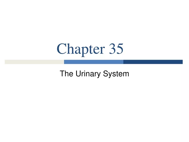
Chapter 35 The Urinary System
Basic Functions • Urinary systems help maintain homeostasis—the relatively constant internal environment • Composition of blood and extracellular fluid • Control the concentration or osmolarity, of dissolved substances in cells and their extracellular environment • Excretion - removal of unwanted substances • Produces urine - contains waste products of cellular metabolism
Urinary System Functions • Three basic processes • Blood or extracellular fluid is filtered, removing water and small dissolved substances • Nutrients are selectively reabsorbed back into the filtered fluid • Excess water, excess nutrients, and dissolved wastes are excreted from the body in urine
Flatworm Urine Systems • The earliest excretory system served to maintain water balance, the primary function of the simple excretory system of flatworms • This system consists of protonephridia - tubules that branch throughout extracellular fluid surrounding flatworm tissues • Collects excess water from the extracellular fluid using ciliated “flame cells” and forces the fluid out through excretory pores • The large body surface of flatworms also serves as an excretory structure through which cellular wastes diffuse
Flatworms Use Protonephridia excretory pore eye spot tubule extracellular fluid cilia nucleus flame cell excretory pore (a) Flatworms use protonephridia
Insect Urinary Systems • Insects have an open circulatory system where hemolymph fills the hemocoel and bathes internal tissues directly • Insect excretory systems are Malpighian tubules that extend outward from the intestine and end blindly within hemolymph • Wastes and nutrients move from the hemolymph into the tubules by diffusion and active transport, water follows by osmosis • Urine is conducted into the intestine, solutes are secreted into the hemolymph by active transport • Produces concentrated urine, which is excreted along with feces
Insects Use Malphigian Tubules abdomen Malpighian tubules intestine hemocoel (filled with hemolymph) rectum cellular and digestive wastes (b) Insects use Malpighian tubules
Earthworm Excretory Systems • In earthworms, mollusks, and other invertebrates, excretion is performed by tubular structures called nephridia • The body cavity is filled with extracellular fluid into which wastes and nutrients diffuse • Each nephridium begins with a funnel-like opening, the nephrostome, ringed with cilia that direct extracellular fluid into a narrow, twisted tubule surrounded by capillaries • As the fluid traverses the tubule, salts and nutrients are reabsorbed back into the capillary blood, leaving the wastes and water behind • Urine is excreted through a nephridiopore • Each segment in an earthworm’s body contains a pair of nephridia
Earthworms Use Nephridia coelom (filled with extracellular fluid) nephridium capillary bed nephrostome nephridiopore (c) Earthworms use nephridia
Vertebrate Urinary Systems • Kidneys - organs of the vertebrate urinary system, where blood is filtered and urine is produced • Because vertebrates live in a wide variety of habitats, vertebrate kidneys face different challenges in maintaining constant conditions within their bodies
Homeostatic Kidney Functions • The mammalian urinary system consists of kidneys, ureters, bladder, urethra • These organs filter the blood, collecting, then excreting dissolved waste products in urine • During filtration, water and dissolved molecules are forced out of the blood • The kidneys return nearly all of the water and nutrients required by the body to the blood • The urine retains wastes, which are expelled
Mammalian Urinary Systems • Helps maintain homeostasis in several ways: • Regulate blood levels of ions - sodium, potassium, chloride, calcium • Maintain proper pH of the blood by regulating hydrogen and bicarbonate ion concentrations • Regulate water content of the blood • Retain important nutrients - glucose and amino acids in the blood • Eliminate cellular waste products as urea • Secrete substances that help regulate blood pressure and oxygen levels
Urea • A waste product of protein digestion • Eliminate nitrogenous wastes that are formed when cells break down amino acids • Nitrogenous wastes from cells enter the blood as ammonia(NH3), toxic • The livers of humans and other mammals convert ammonia into urea, which is less toxic • Urea is filtered from the blood by kidneys and excreted in urine
Urea Formation and Excretion 1 Proteins in food are digested The liver converts ammonia to urea, which is less toxic 4 urea Amino acids are carried in the blood to body cells 2 amino acid Urea is carried in the blood to the kidneys 5 The cells convert the amino groups (-NH2) to ammonia, which is carried in the blood to the liver 3 In kidney nephrons, urea is filtered into the urine 6 ammonia NH3
Human Urinary System • Kidneys - paired organs located on either side of the spinal column, just above the waist • Blood enters each kidney through the renal artery, after blood has been filtered, it exits through the renal vein • Urine leaves the kidney through the ureter - a narrow, muscular tube • Rhythmic contractions of the ureter transports urine to the bladder, a hollow, muscular chamber that collects and stores urine • The bladder wall is lined with smooth muscle and is capable of considerable expansion, accommodating up to a pint of urine
The Human Urinary System left renal artery left kidney left renal vein aorta left ureter vena cava urinary bladder urethra (in penis)
Bladder Sphincters • Urine is contained within the bladder by two sphincter muscles • The internal sphincter, where the bladder joins the urethra, opens automatically during the reflexive contractions of the smooth muscle • The external sphincter, located slightly below the internal sphincter, is under voluntary control, allowing the brain to suppress urination unless the bladder becomes overly full • When open, the sphincters allow urine to flow into the urethra, a single narrow tube that conducts urine outside the body
Kidney Structure • The structure of the kidney supports its function of producing urine • Each kidney contains a solid outer layer - the renal cortex and the inner layer - the renal medulla • The renal medulla surrounds a branched, funnel-like chamber - the renal pelvis, which collects urine and funnels it into the ureter
Cross-Section of a Kidney renal pelvis (cut away to show the path of urine) renal artery renal cortex renal medulla renal pelvis renal vein ureter collecting duct urine to the bladder nephron renal medulla renal cortex enlargement of a single nephron and collecting duct
Nephrons • The renal cortex is made up of more than 1 million microscopic filters or nephrons • Two major parts – • The glomerulus, a dense knot of capillaries where fluid is filtered out of the blood through the porous capillary walls • A long, twisted tubule, where urine formation occurs
Nephron • Bowman’s capsule, a cup-like chamber that surrounds the glomerulus and receives fluid filtered out of the blood from the glomerular capillaries • Collected fluid is conducted to the proximal tubule • The loop of Henle carries the filtered fluid from the cortex, deep into the medulla and back to the cortex • The distal tubule - in the cortex - collects the filtrate from the loop of Henle and passes it to the collecting duct • The collecting duct is not part of the nephron, but collects fluid from many nephrons and deposits it in the renal pelvis
Individual Nephron and Blood Supply collecting duct distal tubule proximal tubule Bowman’s capsule glomerulus arterioles venule branch of the renal artery branch of the renal vein loop of Henle capillaries
The kidney’s blood supply • The kidneys have an enormous blood supply, receiving more than one quart of blood every minute • Blood flows to each kidney from the renal artery, which branches into arterioles that each supply nephrons with blood for filtration • The arterioles branch into capillaries and form the glomerulus of each nephron • The capillaries empty into an outgoing arteriole that branches into capillaries that surround the tubule • The capillaries carry blood into a venule that takes the blood to the renal vein and then the inferior vena cava
Individual Nephron and Blood Supply collecting duct distal tubule proximal tubule Bowman’s capsule glomerulus arterioles venule branch of the renal artery branch of the renal vein loop of Henle capillaries
Urine Production – 3 Stages • Filtration - water and small dissolved molecules are filtered out of the blood • Tubular reabsorption - water and necessary nutrients are restored to the blood • Tubular secretion - wastes and excess ions remaining in the blood are secreted into the urine
Urine Formation and Concentration Filtration: Water, nutrients, and wastes are filtered from the glomerular capillaries into the Bowman’s capsule of the nephron Tubular reabsorption: In the proximal tubule, most water and nutrients are reabsorbed into the blood 2 1 Tubular secretion: Additional wastes are actively transported into the proximal and distal tubules from the blood 3 proximal tubule collecting duct blood leaving the glomerulus distal tubule Bowman’s capsule blood entering the glomerulus loop of Henle Concentration: The loop of Henle produces a salt concentration gradient in the extracellular fluid; in the collecting duct, urine may become more concentrated than the blood as water leaves by osmosis 4
Filtration • Small organic nutrients - amino acids and glucose - are filtered out and returned to the blood • Large quantities of water and ions are filtered out, but the return rate is adjusted to meet changing needs • Ions include sodium (Na+), chloride (Cl–), potassium (K+), calcium (Ca++), hydrogen (H+), and bicarbonate • Urine is formed in the glomerulus and tubule of the nephron • Filtration - when water carrying small dissolved molecules and ions is forced through the walls of the capillaries that form the glomerulus • Blood cells and large proteins are too large to leave the capillaries, so remain in the blood • The fluid filtered out of the glomerular capillaries – the filtrate – collected in Bowman’s capsule and continues through the tubule
Tubular Reaborption • Occurs primarily in the proximal tubule, water and other nutrients are reabsorbed in other tubule areas • Returns organic nutrients - glucose, amino acids, vitamins, ions (Na+, Cl–, K+, Ca2+, H+ and HCO3–) to the blood • Restores most of the water, water follows the nutrients and ions by osmosis through aquaporins - proteins that form water pores • Remaining wastes and excess ions move from the blood into the proximal and distal tubules • Excess K+ and H+, small quantities of ammonia, drugs, food additives, pesticides, and toxins (ie: nicotine) • Tubular secretion occurs primarily by active transport and takes place in both the proximal and distal tubules • When the filtrate leaves the distal tubule, it is urine
The Loop of Henle • Creates an extracellular concentration gradient in the renal medulla • The functions of the loop of Henle • Some water and salt is reabsorbed from the filtrate as it passes through the loop • Most importantly, it creates a high salt and urea concentration in the extracellular fluid within the medulla
Water Regulation • Why is a high salt concentration is important? Water regulation • The kidneys help maintain water content in body tissues by producing…. • dilute, watery urine when fluid intake is high • concentrated urine when fluid intake is low • Water is conserved by allowing it to move out of the collecting duct by osmosis and down its concentration gradient • The more concentrated the extracellular fluid, the more water leaves the urine as it moves through the collecting duct
Summary • The loop of Henle produces and maintains a high salt concentration gradient in the extracellular fluid of the medulla by transporting salt out of the filtrate • The salt and urea gradient causes an osmotic gradient between the filtrate and the surrounding extracellular fluid • The most concentrated fluid surrounds the bottom of the loop • The collecting duct passes through this gradient as it conducts urine from the distal tubule in the renal cortex into the renal pelvis
Details of Urine Formation TUBULAR REABSORPTION & TUBULAR SECRETION URINE CONCENTRATION FILTRATION H+ NH3 some drugs H+ K+ some drugs HCO3– Ca2+ Cl– K+ NaCl Ca2+ Na+ nutrients H2O* H2O 1 H2O* 7 distal tubule proximal tubule 6 2 Bowman’s capsule NaCI H2O renal cortex renal medulla 5 NaCI H2O NaCI 3 H2O 4 (extracellular fluid) urea NaCI 8 H2O* H2O osmosis loop of Henle active transport diffusion collecting duct
Concentration • As the filtrate descends into the loop of Henle and collecting duct… • It is exposed to the osmotic gradient surrounding the nephron • Water leaves the filtrate by osmosis and enters the surrounding capillaries • Filtrate becomes urine once it enters the collecting duct and can be more than four times as concentrated as blood
Kidneys Regulate Osmolarity of Blood • Kidneys regulate the water content of the blood • Human kidneys filter out 1/2 cup of fluid from the blood each minute • Fine-tuning the composition of blood and helping maintain homeostasis • If the kidneys did not reabsorb this water, the rate of filtration would require that we drink 50 gallons of water a day • The urinary system needs to restore nearly all of the water that is initially filtered out of the glomeruli
Antidiuretic Hormone • Antidiuretic hormone (ADH) – Regulates reabsorption and influences the ability of kidneys to reabsorb water • Secreted by the posterior pituitary gland, carried in the blood • It stimulates cells of the distal tubule and collecting ducts to insert more aquaporin proteins into their membranes • The abundance of aquaporin membranes determines the permeability of the membranes to water • Normally some ADH is always present in the blood • Within the hypothalamus, receptors monitor blood osmolarity, which increases when water is lost
An Example • When water is lost during dehydration: • If blood osmolarity exceeds optimal level, the hypothalamus stimulates the pituitary gland to release ADH into the bloodstream • Cells of the distal tubule and collecting duct insert more aquaporins into their membranes, increasing permeability to water • The more concentrated extracellular fluid draws water out by osmosis, restoring water to the blood through nearby capillaries
Dehydration Stimulates ADH Release and Water Retention Heat causes water loss and dehydration through sweating 1 2 Receptors in the hypothalamus detect the increased blood osmolarity and signal the pituitary gland 3 The pituitary gland releases ADH into the bloodstream ADH increases the permeability of the distal tubule and the collecting duct, allowing more water to be reabsorbed into the blood 4 Water is retained in the body and concentrated urine is produced 5
Kidneys regulate BP and Oxygen • Kidneys release substances that help regulate blood pressure and oxygen levels • When blood pressure falls, kidneys release renin • Renin catalyzes the formation of the hormone angiotensin in the blood
Angiotensin • Combats low blood pressure in three ways • It stimulates the proximal tubules of nephrons to reabsorb more Na+ into the blood, causing water to follow by osmosis (increase blood volume) • It stimulates ADH release, causing more water to be reclaimed from the distal tubule and collecting duct • It causes arterioles throughout the body to constrict, which directly increases blood pressure
Erythropoietin • When blood oxygen levels are low, kidneys release erythropoietin • Stimulates the bone marrow to make more red blood cells • The higher number of red blood cells increases the oxygen carrying capacity of the blood
Vertebrate kidneys are adapted to diverse environments • Mammals have structurally different nephrons, depending upon the availability of water in their natural habitat • Mammals adapted to dry climates have long loops of Henle • Longer loops allow a higher concentration of salt to be produced in the extracellular fluid of the medulla, so more water is reclaimed from the collecting duct • A mammal with very long-looped nephrons is the desert kangaroo rat
More Mammal Adaptations • Mammals adapted to habitats with an abundance of fresh water have short loops of Henle • Beavers, which live along streams, can only concentrate their urine to about twice their blood osmolarity • Humans have a mixture of long- and short-looped nephrons, and can concentrate urine up to four times the osmolarity of blood
Freshwater Fish • Animals have evolved homeostatic mechanisms, including kidney adaptations, to maintain water and salt within their bodies – osmoregulation • Freshwater fish live in a hypotonic environment • Water continuously moves into their bodies by osmosis, salts diffuse out • Freshwater fish acquire salt from food and through their gills but never drink • Their kidneys retain salt and excrete large quantities of extremely dilute urine
Osmoregulation in Fish fresh water water salt Water moves in by osmosis; salt diffuses out Salt is pumped in by active transport Salt and some water enters in food The kidneys conserve salt and excrete large amounts of dilute urine (a) Freshwater fish
Saltwater Fish • Saltwater fish live in a hypertonic environment; seawater has a solute concentration of two to three times that of their body fluids • Water is constantly leaving their tissues by osmosis, and salt is constantly diffusing in and being taken in with food • To compensate, saltwater fish drink to restore their lost water, and excess salt they take in is excreted by active transport through their gills
Osmoregulation in Fish salt water Salt and water enter in food and by drinking seawater Water moves out by osmosis; salt diffuses in Salt is pumped out by active transport Some salt is excreted in small quantities of urine water salt (b) Saltwater fish