Download
1 / 10
100 likes | 248 Views
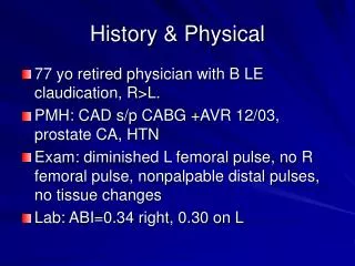
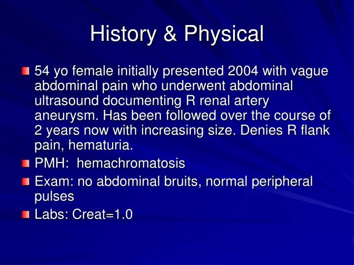
History & Physical. 54 yo female initially presented 2004 with vague abdominal pain who underwent abdominal ultrasound documenting R renal artery aneurysm. Has been followed over the course of 2 years now with increasing size. Denies R flank pain, hematuria. PMH: hemachromatosis

E N D
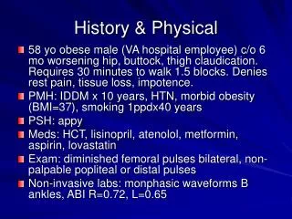
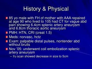
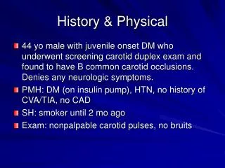
History & Physical • 54 yo female initially presented 2004 with vague abdominal pain who underwent abdominal ultrasound documenting R renal artery aneurysm. Has been followed over the course of 2 years now with increasing size. Denies R flank pain, hematuria. • PMH: hemachromatosis • Exam: no abdominal bruits, normal peripheral pulses • Labs: Creat=1.0
R kidney removed from Gerota’s fascia Ureter dissected free
Kocher maneuver frees up IVC IVC R ovarian vein ligated R renal vein Ureter dissected free
ureter After artery and vein clamped and ligated off aorta and IVC, kidney brought up to ice bucket Bifurcation of main R renal artery with thin walled-aneurysm
R main renal artery Renal vein transected
Superior & inferior renal arteries transected, then syndactylized
Syndactylized branches R renal artery R hypogastric artery