Download
1 / 122
1.84k likes | 3.76k Views
Biomechanics of Bone. Prepared by: Dr. Faryal Zaidi. 1)BRIEF REVIEW OF BONE BIOLOGY, STRUCTURE, AND CHEMICAL COMPOSITION 2) MECHANICAL PROPERTIES OF BONE Material Properties versus Geometry Anisotropy Elastic Constants of Bone Strength Fracture Toughness Strain Rate

E N D
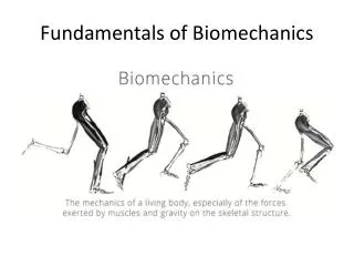
Biomechanics of Bone Prepared by: Dr. Faryal Zaidi
1)BRIEF REVIEW OF BONE BIOLOGY, STRUCTURE, AND CHEMICAL COMPOSITION 2) MECHANICAL PROPERTIES OF BONE Material Properties versus Geometry • Anisotropy • Elastic Constants of Bone • Strength • Fracture Toughness • Strain Rate 3) CHANGES IN MECHANICAL PROPERTIES WITH AGE AND ACTIVITY 4) FRACTURES
The purposes of this chapter are to • Briefly review basic bone biology and terminology • Describe mechanical properties of human bone • Discuss the clinical relevance of understanding bone properties
Overall Composition of Bone • Two major components • Organic matrix • Type I collagen • Amorphous ground substance • Inorganic matrix • Calcium hydroxyapatitie.
Organic Matrix Organic matter (40% of dry weight of bone) • Consisting of type I collagen fibresembedded in the ground substance • containing proteoglycans and glycoproteins.The collagen fibres are made up of bundles of fibrils to resist pulling forces.
Organic Matrix • 40% dry weight • collagen (90%) • proteoglycans • non-collagenous matrix proteins • growth factors • cytokines
Inorganic matter (60% of dry weight of bone) • Made up of stiffening substancesto resist bending and compression. • The bone mineral is an analogue of crystals of calcium phosphate —hydroxyapatiteCa10(PO4)6(OH)2. It is this association of hydroxyapatite with collagen fibres which is responsible for the hardness of bone. • Provides the compressive strength of bone • Responsible for the mineralization of bone
Histological Types of Bones Bone as a tissue consists of two main types: Primary bone tissue (non-lamellar bone) • This bone is also known as ‘coarse fibred’ or ‘woven’ bone or immature bone. It is characterized by the presence of randomly distributed coarse collagen fibres. • eg foetuses and young children.
Woven bone • It is also the first tissue to appear in the repair of bone (fracture healing). • The random distribution of fibers gives some strength in all directions, but woven bone is not as strong as mature bone. • It is also more flexible than adult bone, presumably to provide resilience for all the falls and tumbles of childhood.
Lamellar bone Secondary bone tissue (lamellar bone) • known as mature bone. characterised by the presence of collagen fibres arranged in parallel layers or sheets (lamellae). • Lamellar bone is present in both structured types of adult bone, cortical (compact) bone and cancellous (spongy or trabecular) bone.
Running perpendicular to the haversian canals are Volkmann’s canals. They connect the blood and nerve supply in the periosteum to those in the haversian canals and the medullary cavity.
Cortical Bone • Cortical or compact bone is made up of a structure of Haversian systems or Osteons. • Each Osteon is a cylinder running parallel to the long axis of the diaphysis. • In the centre of each osteon is the Haversian canal which is lined by endosteum containing blood vessels, nerves and loose connective tissue. • Surrounding each canal are 4–20 concentric lamellae of collagen fibres. The Haversian canals are round or oval in cross-section. They generally run in a longitudinal direction.
80% skeleton • Cement lines define outer border of osteon • Slow turnover • Relatively high Young’s modulus(stress/strain) • Higher resistance to torsion and bending than cancellous.
All the collagen fibers in a particular lamella run in a single direction, while collagen fibers in adjacent lamellae will run in the opposite direction. This allows bone to better withstand twisting forces.
Fig. 4: Compact bone consisting of cylindrical units — Osteons, each Osteon with aHaversian canal in its centre and surrounded by a cement line. In between the Osteons are the interstitial lamellae.
Each osteon communicates with the marrow cavity, the periosteum and with each other through transverse or oblique canals – the Volkmann’s canals. • The osteocytes are arranged circumferentially around the central canal in parallel with the lamellae and are interconnected by fine processes of osteocyte cytoplasm—the filopodia. • The osteocytes are housed in lacunae interconnected by canaliculi containing these fine cytoplasmic processes.
Running perpendicular to the haversian canals are Volkmann’s canals. They connect the blood and nerve supply in the periosteum to those in the haversian canals and the medullary cavity.
Lying in between intact osteons are incomplete lamellae called interstitial lamellae. These fill the gaps between osteons or are remnants of bone remodeling. (remnants of previous Haversian systems which have been remodelled) There are also circumferential lamellae that extend around the circumference of the shaft. There are inner circumferential lamellae surrounding the endosteum and outer circumferential lamellae just inside the periosteum.
Each osteon is separated from its neighbour and from interstial lamellae by a cement line.
Cancellous Bone • Less dense • More remodeling along lines of stress (Wolff’s law) • Much larger surface area • Higher turnover • Lower apparent modulus • More elastic • More resistance to compressive forces
Cancellous Bone • Cancellous or spongy bone consists of a series of interconnecting plates of bone — the trabeculae. Each bone trabecula contains collagen fibres arranged in parallel lamellae. • The surface of the trabecula is covered by an attenuated layer of flattened cells, the resting osteoblasts.
Such a structure, in addition to providing a large surface area for metabolic activities of bone, gives mechanical strength without the disadvantages of undue weight. • The thickest and strongest trabeculae are arranged in the direction subjected to the greatest stress (Wolff’s Law).
Trabeculae are a few cell layers thick and contain irregularly arranged lamellae and osteocytes interconnected by canaliculi. • No haversian or Volkmann’s canals are necessary. Why?
Wolff’s Law http://www.bartleby.com/107/Images/small/image247.jpg
Bone Remodeling • Bone is a dynamic tissue. • What does that mean? • Wolff’s law holds that bone will grow or remodel in response to the forces or demands placed on it. Examine this with the bone on the left.
Bone Remodeling • Bone is a dynamic tissue. • What does that mean? • Wolff’s law holds that bone will grow or remodel in response to the forces or demands placed on it. Examine this with the bone on the left.
Wolff's law states that bones develop a structure most suited to resist the forces acting upon them, adapting both the internal architecture and the external conformation to the change in external loading conditions.
The internal architecture is adapted in terms of change in density and in disposition of trabecules and osteons and the external conformation in terms of shape and dimensions. When strain is intensified new bone is formed.
Formation and remodelling Bone formation is an essential process in the development of the human body. It starts during the development of the foetus, and continues throughout childhood and adolescence as the skeleton grows. Bone remodelling meanwhile is a life-long process, consisting of resorption (the breaking down of old bone) and ossification (formation of new bone), and is key to shaping the skeleton and to the repair of bone fractures.
There are three types of cell present in bone that are of particular interest – • osteoblasts, • osteocytes • osteoclasts, which are respectively responsible for the production, maintenance and resorption of bone.
Osteoblasts Mononucleated “bone-forming” cells found near the surface of bones. They are responsible for making osteoid, which consists mainly of collagen. The osteoblasts then secrete alkaline phosphatase to create sites for calcium and phosphate deposition, which allows crystals of bone mineral to grow at these sites. The osteoid becomes mineralised, thus forming bone.
Osteocytes These are osteoblasts that are no longer on the surface of the bone, but are instead found in lacunae between the lamellae in bone. Their main role is homeostasis – maintaining the correct oxygen and mineral levels in the bone.
Osteoclasts Multinucleated cells responsible for bone resorption. They travel to specific sites on the surface of bone and secrete acid phosphatase, which unfixes the calcium in mineralised bone to break it down.
Bone resorption Bone deposition
Bone resorption and bone deposition processes are always active in bone. • An equilibrium strain state exists in correspondence to which the two activities are perfectly balanced. • When strain intensity is higher than the equilibrium strain deposition activity is more intense than resorption activity and net deposition occurs.
When strain intensity is lower than the equilibrium strain deposition activity is less intense than resorption activity and net resorption occurs. • Dynamical equilibrium between resorption and deposition is again achieved when the equilibrium strain state is newly established.
Bone has the ability to remodel, by altering its size, shape, and structure, to meet the mechanical demands placed on it (Buckwalter et al., 1995). • Wolff's law states that the remodeling of bone is influenced and modulated by mechanical stresses (Wolff, 1892).
Examples are The racquet-holding arm bones of tennis players become much stronger than those of the other arm. Their bodies have strengthened the bones in their racquet-holding arm since it is routinely placed under higher than normal stresses. Weightlifters often display increases in bone density in response to their training
Load on the skeleton can be accomplished by either muscle activity or gravity. • A positive correlation exists between bone mass and body weight. A greater body weight has been associated with a larger bone mass (Exner et al., 1979).
Conversely, a prolonged condition of weightlessness, such as that experienced during space travel, has been found to result in decreased bone mass in weight-bearing bones. • These changes are not completely reversible.
Disuse or inactivity has deleterious effects on the skeleton. Bed rest induces a bone mass decrease of approximately 1% per week (Jenkins & Cochran, 1969; Krolner & Toft, 1983).
An implant that remains firmly attached to a bone after a fracture has healed may also diminish the strength and stiffness of the bone. • In the case of a plate fixed to the bone with screws, the plate and the bone share the load in proportions determined by the geometry and material properties of each structure.