Download
1 / 19
190 likes | 366 Views
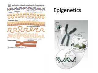
V2 epigenetics during development. Review of lecture V1 ... What is: - bisulfite treatment of DNA - methylation-specific PCR - Restriction landmark genomic scanning (RLGS) - chromatin immunoprecipitation using the ChIP-on-chip approach?. epigenetics during specialisation of cells.

E N D
V2 epigenetics during development Review of lecture V1 ... What is: - bisulfite treatment of DNA - methylation-specific PCR - Restriction landmark genomic scanning (RLGS) - chromatin immunoprecipitation using the ChIP-on-chip approach? Biological Sequence Analysis
epigenetics during specialisation of cells Key feature of multi-cellular organisms: ability to develop specialized cells with specific functions. Process depends on cellular differentiation pathways. Initiation of such pathways is determined by coordinated regulation of; - silent genes that in many cases have never been expressed must be activated, - a number of transcriptionally competent genes must be repressed. Essential in understanding differentiation: identify tissue-specific genes and regulatory proteins that directly control their expression. de la Serna et al. Nat. Rev. Gen. 7, 461 (2006) Biological Sequence Analysis
specialisation of cells However, tissue-specific transcriptional regulatory proteins are not sufficient to initiate differentiation. Also essential: - changes at the level of both higher-order chromatin structure and - chromatin organization at individual genes. Proteins are needed that alter the structure of chromatin at lineage-specific genes to facilitate the function of tissue-specific regulators. de la Serna et al. Nat. Rev. Gen. 7, 461 (2006) Biological Sequence Analysis
Chromatin-remodelling enzymes • Two main classes: • - enzymes that covalently modify histone proteins (see V1), and • - enzymes that use ATP hydrolysis to alter histone–DNA contacts. • Both classes have significant roles in gene regulation, • including differentiation-specific gene expression. • ATP-dependent remodellers don‘t function similarly in all cell types. • Instead, they have a range of specific and context-dependent roles in differentiation. E.g. they have functions in • - recombination, • - cell-cycle regulation and • genome organization, • indicating important links between chromatin remodelling and other cellular processes during differentiation. de la Serna et al. Nat. Rev. Gen. 7, 461 (2006) Biological Sequence Analysis
ATP-dependent chromatin-remodelling enzymes The three best-characterized classes of ATP-dependent chromatin-remodelling enzyme are the families of - SWI/SNF, - CHD (chromodomain and helicase-like domain) and - ISWI (imitation SWI). Each has a unique domain (bromo, chromo and sant) that likely interact with specific chromatin substrates. Each enzyme class forms complexes with other proteins: - SWI/SNF proteins interact with brahma (BRM)- or brahma-like 1 (BRG1)-containing enzymes. - CHD proteins can form part of the NuRD (nucleosome remodelling and histone deacetylase) complex, which can include CHD3- or CHD4-containing enzymes, or possibly both. - ISWI SNF2H-containing enzymes are found in several complexes (for example, ACF (ATPutilizing chromatin assembly and remodelling factor) and RSF (remodelling and spacing factor)), and SNF2L enzymes form part of the NuRF (nucleosome-remodelling factor) and CERF (CECR2-containing remodelling factor) complexes. de la Serna et al. Nat. Rev. Gen. 7, 461 (2006) Biological Sequence Analysis
Example: skeletal muscle differentiation • The myogenin gene (Myog) is expressed specifically during skeletal muscle differentiation. The locus is constitutively bound by a heterodimer of 2 homeodomain proteins from the PBX/MEIS family in undifferentiated cells. • E : binding sites for the transcription factor MyoD; • P : binding sites for the transcription factor PBX; • M : binding sites for the transcription factor MEF2; • T : the TATA box for Myog. • In undifferentiated cells, several of these sites are inaccessible to the proteins that bind them due to the conformation of chromatin at this locus (indicated by crosses). • Initial targeting of the skeletal muscle regulator, MyoD, to the myogenin promoter occurs in part through physical interactions with PBX. • MyoD then sequentially targets • histone acetyl transferase (HAT) enzymes — which acetylate (Ac) both promoter histones and MyoD — and • a BRG1-based SWI/SNF enzyme, which is activated through the p38 kinase-mediated phosphorylation (yellow circle) of the BAF60 subunit. • The SWI/SNF enzyme mediates ATP-dependent chromatin remodelling at the myogenin promoter, which results in changes in accessibility that permit the stable binding of heterodimers of MyoD and an E-box binding protein (EBP), and another factor, MEF2, to their cognate binding sites in the myogenin promoter. • Then transcription of Myog can take place. de la Serna et al. Nat. Rev. Gen. 7, 461 (2006) Biological Sequence Analysis
Abstract: A unique feature of the germ cell lineage is the generation of totipotency. A critical event in this context is DNA demethylation and the erasure of parental imprints in mouse primordial germ cells (PGCs) on embryonic day 11.5 (E11.5) after they enter into the developing gonads. Little is yet known about the mechanism involved, except that it is apparently an active process. We have examined the associated changes in the chromatin to gain further insights into this reprogramming event. Here we show that the chromatin changes occur in two steps. The first changes in nascent PGCs at E8.5 establish a distinctive chromatin signature that is reminiscent of pluripotency. Next, when PGCs are residing in the gonads, major changes occur in nuclear architecture accompanied by an extensive erasure of several histone modifications and exchange of histone variants. Furthermore, the histone chaperones HIRA and NAP-1 (NAP111), which are implicated in histone exchange, accumulate in PGC nuclei undergoing reprogramming. We therefore suggest that the mechanism of histone replacement is critical for these chromatin rearrangements to occur. The marked chromatin changes are intimately linked with genome-wide DNA demethylation. On the basis of the timing of the observed events, we propose that if DNA demethylation entails a DNA repair-based mechanism, the evident histone replacement would represent a repair-induced response event rather than being a prerequisite. Biological Sequence Analysis
some terms from developmental biology somatic cells = cells forming the body of an organism germ cells (dt. Keimzelle, Ovolum) are part of the germline. germline (dt. Keimbahn)= line of germ cells that have genetic material that may be passed to a child/embryo. Germline cells are immortal. Gametocyte = eukaryotic germ cell; includes spermatocytes (male) and oocytes (female) primordial germ cells : predecessors of germ cells. They migrate to the gonadal ridge. They may be detected from expression of Stella gonad (dt. Keimdrüse) gonadal ridge = precursor to the gonads www.wikipedia.org Biological Sequence Analysis
Germ line development Germline cells are produced by embryonic cleavage. Cleavage: division of cells in the early embryo. The zygotes of many species undergo rapid cell cycles with no significant growth. The different cells derived from cleavage are called blastomeres. Cleavage in mammals is slow. Cell division takes 12 – 24 hours and is asynchronous. In mammals, specification of germ cells seems to proceed by induction. BMP (Bone morphogenetic protein) signals from the extraembryonic ectoderm activate expression of fragilis and bias the cells toward PGC. The cells expressing fragilis collectively express stella and Blimp1, a general repressor of transcription. www.wikipedia.org Biological Sequence Analysis
Working hypothesis • The specification of about 40 primordial germ cells (PGCs) from Blimp1-expressing PGCs precursors is accompanied by expression of stella on E7.25. • After their migration into the developing gonads, PGCs show genome-wide DNA demethylationbetween E11.5 and E12.5, including erasure of genomic imprints, which is supposedly an active process. • The mechanism of this DNA demethylation process is unknown, but we reasoned that it might be linked with changes in chromatin and histone modifications. • Investigate chromatin in nascent PGCs at E8.5 (100 PGCs per embryo) Hajkova et al. Nature 452, 877 (2008) Biological Sequence Analysis
PGC development in mouse The nascent (dt. im Entstehen begriffenen) PGCs are first identified on E7.5 as a group of about 40 stella expressing cells. On E8.5 (Step1 of the reprogramming process) when there are about 1000 PGCs per embryo, they start to migrate along the developing hindgut (dt. Dickdarm) and reach the developing gonads at about E10.5. Soon after the entry into the gonads the PGCs undergo epigenetic reprogramming (as a second step of the reprogramming process), which includes genome-wide DNA demethylation, erasure of genomic imprints and re-activation of the inactive X chromosome (Xi) in female embryos. Hajkova et al. Nature 452, 877 (2008) Biological Sequence Analysis
chromatin changes - loss of dimethylation of Lys9 of histone H3 (H3K9me2) - in early PGCs enhanced trimethylation of H3K27me3 - enriched methylation of H3K4me2 and H3K4me3 and of many histone acetylation marks, especially H3K9ac, as well as symmetrical methylation of Arg3 on histones H4 and H2A (H4/H2AR3me2s). Notably, this germ cell chromatin signature is established specifically in PGCs (not detected in the contemporary somatic cells) before their entry into the gonads, and is associated with the expression of pluripotency-specific genes: Sox2, Oct4(Pou5f1), Nanog and stella. What are these 4 genes: Sox2, Oct4, Nanog, stella? Hajkova et al. Nature 452, 877 (2008) Biological Sequence Analysis
Sox2 = SRY (sex determining region Y)-box 2 SRY or SOX2 is a transcription factor that is essential to maintain self-renewal of undifferentiated embryonic stem cells. Intronless gene. The encoded protein may act as a transcriptional activator after forming a protein complex with other proteins. www.wikipedia.org http://symatlas.gnf.org/SymAtlas/ Biological Sequence Analysis
Oct4 (ASH1L, POU5F1) Oct4 is expressed in developing embryos throughout the preimplantation period. Knockout of Oct-4 promotes differentiation. One of its main functions is to keep the embryo from differentiating. Too much or too little expression will cause differentiation of the cells. Therefore, it is frequently used as a marker for undifferentiated cells. http://symatlas.gnf.org/SymAtlas/ www.wikipedia.org Biological Sequence Analysis
Nanog Nanog is another gene expressed in embryonic stem cells (ESC) and is thought to be a key factor in maintaining pluripotency. Nanog works together with other factors as POU5F1 and SOX2 to establish ESC identity. Human nanog: 305 amino acid protein, conserved homeodomain that facilitates DNA binding. http://symatlas.gnf.org/SymAtlas/ www.wikipedia.org Biological Sequence Analysis
PGC development in mouse Scheme summarising chromatin changes occurring in PGCs during the reprogramming process. The chromatin changes in PGCs occur in 2 steps. The first step is characterized by loss of H3K2me2 and gain of H3K27me3, H3K9ac and H4/H2A R3me2s at E8.5. The second step occurs at E11.5 and is characterized by changes in nuclear architecture (loss of chromocenters) and by the loss of numerous histone modifications. Hajkova et al. Nature 452, 877 (2008) Biological Sequence Analysis
DAPI staining: labelling of cell nucleus DAPI: 4‘,6-diamidino-2-phenylindole is a fluorescent stain that binds strongly to DNA. It can pass through intact cell membranes. When bound to ds-DNA, DAPI absorbs maximally at 358 nm and emits fluorescent light at 461 nm (blue/cyan). Hajkova et al. Nature 452, 877 (2008) Biological Sequence Analysis
methylation of CpG islands What does the differential methylation mean? Hajkova et al. Nature 452, 877 (2008) Biological Sequence Analysis
connections between chromatin and DNA methylation - What do the two models describe? - How did the authors arrive at the two models? - How could one distinguish between these two models? Hajkova et al. Nature 452, 877 (2008) Biological Sequence Analysis