Download
1 / 1
10 likes | 64 Views
GSKIP, shares a homology of Axin GID domain and functions as a negative regulator of GSK3beta

E N D
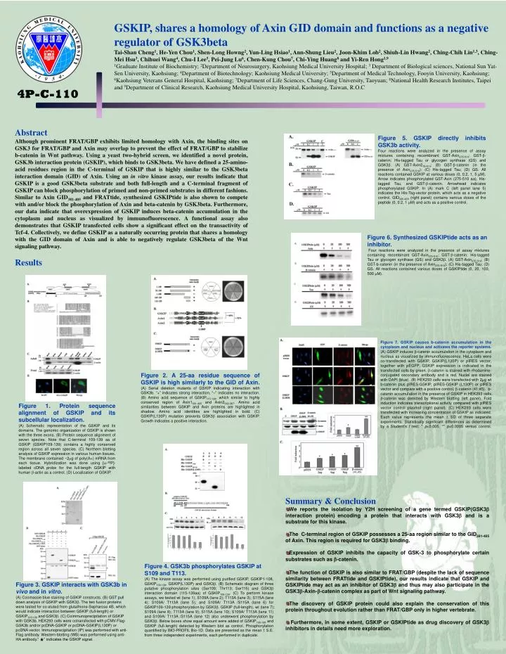
GSKIP, shares a homology of Axin GID domain and functions as a negative regulator of GSK3beta Tai-Shan Cheng1, He-Yen Chou1, Shen-Long Howng2, Yun-Ling Hsiao1, Ann-Shung Lieu2, Joon-Khim Loh2, Shiuh-Lin Hwang2, Ching-Chih Lin1,3, Ching-Mei Hsu3, Chihuei Wang4, Chu-I Lee5, Pei-Jung Lu6, Chen-Kung Chou7, Chi-Ying Huang8 and Yi-Ren Hong1,9 1Graduate Institute of Biochemistry; 2Department of Neurosurgery, Kaohsiung Medical University Hospital; 3 Department of Biological sciences, National Sun Yat-Sen University, Kaohsiung; 4Department of Biotechnology; Kaohsiung Medical University; 5Department of Medical Technology, Fooyin University, Kaohsiung; 6Kaohsiung Veterans General Hospital, Kaohsiung; 7Department of Life Sciences, Chang-Gung University, Taoyuan; 8National Health Research Institutes, Taipei and 9Department of Clinical Research, Kaohsiung Medical University Hospital, Kaohsiung, Taiwan, R.O.C 4P-C-110 Abstract Although prominent FRAT/GBP exhibits limited homology with Axin, the binding sites on GSK3 for FRAT/GBP and Axin may overlap to prevent the effect of FRAT/GBP to stabilize b-catenin in Wnt pathway. Using a yeast two-hybrid screen, we identified a novel protein, GSK3b interaction protein (GSKIP), which binds to GSK3beta. We have defined a 25-amino-acid residues region in the C-terminal of GSKIP that is highly similar to the GSK3beta interaction domain (GID) of Axin. Using an in vitro kinase assay, our results indicate that GSKIP is a good GSK3beta substrate and both full-length and a C-terminal fragment of GSKIP can block phosphorylation of primed and non-primed substrates in different fashions. Similar to Axin GID381-405 and FRATtide, synthesized GSKIPtide is also shown to compete with and/or block the phosphorylation of Axin and beta-catenin by GSK3beta. Furthermore, our data indicate that overexpression of GSKIP induces beta-catenin accumulation in the cytoplasm and nucleus as visualized by immunofluorescence. A functional assay also demonstrates that GSKIP transfected cells show a significant effect on the transactivity of Tcf-4. Collectively, we define GSKIP as a naturally occurring protein that shares a homology with the GID domain of Axin and is able to negatively regulate GSK3beta of the Wnt signaling pathway. Figure 5. GSKIP directly inhibits GSK3b activity. Four reactions were analyzed in the presence of assay mixtures containing recombinant GST-Axin275-510; GST-b-catenin; His-tagged Tau or glycogen synthase (GS) and GSK3b. (A) GST-Axin275-510; (B) GST-b-catenin (in the presence of Axin275-510); (C) His-tagged Tau; (D) GS. All reactions contained GSKIP at various doses (0, 0.2, 1, 5 µM). Arrow indicates phosphorylated GST-Axin (275-510 aa), His-tagged Tau, and GST-b-catenin. Arrowhead indicates phosphorylated GSKIP. In (A) mark C (left panel lane 5) indicates the His-Tag-vector protein, which acts as a negative control. GID320-429 (right panel) contains various doses of the peptide (0, 0.2, 1 mM) and acts as a positive control. Figure 6. Synthesized GSKIPtide acts as an inhibitor. Four reactions were analyzed in the presence of assay mixtures containing recombinant GST-Axin275-510; GST-b-catenin; His-tagged Tau or glycogen synthase (GS) and GSK3b. (A) GST-Axin275-510; (B) GST-b-catenin (in the presence of Axin275-510); (C) His-tagged Tau; (D) GS. All reactions contained various doses of GSKIPtide (0, 20, 100, 500 mM). Results Figure 7. GSKIP causes b-catenin accumulation in the cytoplasm and nucleus and activates the reporter systems. (A) GSKIP induces b-catenin accumulation in the cytoplasm and nucleus as visualized by immunofluorescence. HeLa cells were co-transfected with GSKIP, GSKIP(L130P) or pIRES vector, together with pEGFP. GSKIP expression is indicated in the transfected cells by green. b-catenin is stained with rhodamine-conjugated secondary antibody and is red. Nuclei are stained with DAPI (blue). (B) HEK293 cells were transfected with 2µg wt b-catenin plus pIRES-GSKIP, pIRES-GSKIP (L130P) or pIRES vector and compare with a positive control, b-catenin (41;45). b-catenin accumulation in the presence of GSKIP in HEK293 cells. b-catenin was detected by Western blotting (left panel). Fold induction indicates transcriptional activity compared with pIRES vector control plasmid (right panel). (C) HEK293 cells were transfected with increasing concentration of GSKIP as indicated. Each value represents the mean ± SD of three separate experiments. Statistically significant differences as determined by a Student’s t test: * p<0.005, ** p<0.0005 versus control. Figure 2. A 25-aa residue sequence of GSKIP is high similarly to the GID of Axin. (A) Serial deletion mutants of GSKIP indicating interaction with GSK3b. “+” indicates strong interaction, “–” indicates no interaction. (B) Amino acid sequence of GSKIP115-139, which similar to highly conserved region of Axin1381-405 and Axin2363-387. Amino acid similarities between GSKIP and Axin proteins are highlighted in shadow. Amino acid identities are highlighted in bold. (C) GSKIP(L130P) mutation prevents GSK3b association with GSKIP. Growth indicates a positive interaction. Figure 1. Protein sequence alignment of GSKIP and its subcellular localization. (A) Schematic representation of the GSKIP and its domains. The genomic organization of GSKIP is shown with the three exons. (B) Protein sequence alignment of seven species. Note that C-terminal 109-139 aa of GSKIP (GSKIP109-139) contains a highly conserved region across all seven species. (C) Northern blotting analysis of GSKIP expression in various human tissues. The membrane contained ~2mg of poly(A+) mRNA from each tissue. Hybridization was done using [a-32P]-labeled cDNA probe for the full-length GSKIP with human b-actin as a control. (D) Localization of GSKIP. • Summary & Conclusion • We reports the isolation by Y2H screening of a gene termed GSKIP(GSK3b interaction protein)encoding a protein that interacts with GSK3b and is a substrate for this kinase. • The C-terminal region of GSKIP possesses a 25-aa region similar to the GID381-405 of Axin. This region is required for GSK3b binding. • Expression of GSKIP inhibits the capacity of GSK-3 to phosphorylate certain substrates such as b-catenin. • The function of GSKIP is also similar to FRAT/GBP (despite the lack of sequence similarity between FRATtide and GSKIPtide), our results indicate that GSKIP and GSKIPtide may act as an inhibitor of GSK3b and thus may also participate in the GSK3b-Axin-b-catenin complex as part of Wnt signaling pathway. • The discovery of GSKIP protein could also explain the conservation of this protein throughout evolution rather than FRAT/GBP only in higher vertebrate. • Furthermore, in some extent, GSKIP or GSKIPtide as drug discovery of GSK3b inhibitors in details need more exploration. Figure 4. GSK3b phosphorylates GSKIP at S109 and T113. (A) The kinase assay was performed using purified GSKIP, GSKIP1-108, GSKIP109-139, GSKIP(L130P) and GSK3b. (B) Schematic diagram of three putative phosphorylation sites (Ser109; Thr113; Ser115) and GSK3b interaction domain (115-139aa) of GSKIP109-139. (C) To perform kinase assays, we tested wt (lane 1); S109A (lane 2); T113A (lane 3); S115A (lane 4); S109A/ T113A (lane 5); and S109A/ T113A /S115A (lane 6) for GSKIP109-139 phosphorylation by GSK3b. GSKIP (full-length), wt (lane 7); S109A (lane 8); T113A (lane 9); S115A (lane 10); S109A/ T113A (lane 11) and S109A/ T113A /S115A (lane 12) also underwent phosphorylation by GSK3b. Below boxes show equal amount were added of GSKIP109-139 and GSKIP (full-length) detected by Western blot as control. Phosphorylation quantified by BIO-PROFIL Bio-1D. Data are presented as the mean ± S.E. from three independent experiments, each performed in duplicate. Figure 3. GSKIP interacts with GSK3b in vivo and in vitro. (A) Coomassie blue staining of GSKIP constructs. (B) GST pull down analysis of GSKIP with GSK3b. The two fusion proteins were tested for co-eluted from glutathione-Sepharose 4B, which would indicate interaction between GSKIP (full-length) or GSKIP109-139 and GSK3b. (C) Coimmunoprecipitation of GSKIP with GSK3b. HEK293 cells were cotransfected with pCMV-Flag-GSK3b and/or pcDNA-GSKIP or pcDNA-GSKIP(L130P) or pcDNA vector. Immunoprecipitation (IP) was performed with anti-Flag antibody. Western-blotting (WB) was performed using anti-HA antibody.“ ★” indicates the GSKIP signal.