Download
1 / 39
430 likes | 874 Views
SURGICAL ANATOMY OF THE NASOPHARYNX. Dr. Supreet Singh Nayyar, AFMC For more presentations, visit www.nayyarENT.com. Layout. General Description Layers Of The Nasopharyngeal Wall Fascial Relations Of The Nasopharynx Muscles Of The Nasopharynx Blood Vessels Of The Nasopharynx

E N D
SURGICAL ANATOMY OF THE NASOPHARYNX Dr. Supreet Singh Nayyar, AFMC For more presentations, visit www.nayyarENT.com www.nayyarENT.com
Layout • General Description • Layers Of The Nasopharyngeal Wall • Fascial Relations Of The Nasopharynx • Muscles Of The Nasopharynx • Blood Vessels Of The Nasopharynx • Lymphatics Of The Nasopharynx • Nerves Of The Nasopharynx • Imaging Of The Nasopharynx www.nayyarENT.com
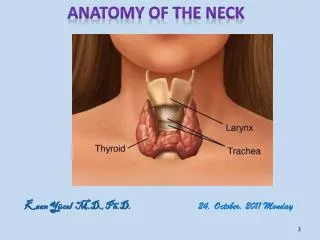
Development Dimensions Boundaries Anterior wall The Floor The roof and posterior wall The lateral wall General Description www.nayyarENT.com
Fossa of Rosenmuller • Location- • Depth- 2.5 cm • Anatomical relationship www.nayyarENT.com
LAYERS OF THE NASOPHARYNGEAL WALL • The mucosa - three types of epithelium • Lymphoid nodules – Waldeyer’s ring • The submucosa • The muscular layer -outer circular & inner longitudinal muscle • The buccopharyngeal fascia www.nayyarENT.com
FASCIAL RELATIONS OF THE NASOPHARYNX • Cervical fascial layers superficial and deep cervical fascia • Cervical fascial spaces midline spaces: pharyngeal mucosal, retropharyngeal and prevertebral spaces Paired lateral spaces: parapharyngeal, carotid, masticator and parotid spaces www.nayyarENT.com
Cervical fascial layers • Superficial cervical fascia -Envelops platysma and muscles of facial expression • Deep cervical fascia - Superficial layer - middle layer - deep layer www.nayyarENT.com
Pharyngeal mucosal space • Encloses the pharynx • Buccopharyngeal fascia • Sinus of morgagni • Buccopharyngeal fascia fuses www.nayyarENT.com
Retropharyngeal space • Location: buccopharyngeal fascia and prevertebral fascia • Extent: skull base to T2 • Two compartments • Source of Infection: extension from para pharyngeal, masticator, parotid space www.nayyarENT.com
Prevertebral space • Location: posterior to the prevertebral fascia • Extent: skull base to coccyx • Source of Infection: TB spine , penetrating trauma www.nayyarENT.com
Carotid space • Extent: jugular foramen to the aortic arch • Location: lateral to the prevertebral www.nayyarENT.com
Masticator space • Extent: skull base to lower border mandible • Location: superficial layer of deep cervical fascia • Source of Infection: 3rd molar www.nayyarENT.com
Parotid space • Location: lateral to the parapharyngeal space, anterior to carotid space and posterior to the masticator space • Extent: superficial layer of deep cervical fascia • Source of Infection: oral cavity via Stenson’sduct www.nayyarENT.com
Contents of the cervical fascial spaces • Pharyngeal mucosal space: mucosa, lymphoid tissue, muscles of pharynx, minor salivary glands • Retropharyngeal space: fat, lymph nodes • Prevertebral space: vertebrae and prevertebral muscles www.nayyarENT.com
Para pharyngeal space: fat, arteries veins, trigeminal nerve, salivary gland rests, lymph nodes • Carotid space: carotid artery, internal jugular vein, cranial nerves IX-XII lymph nodes, Sympathetic fibers • Masticator space: mandible, muscles trigeminal nerve • Parotid space: parotid gland, facial nerve, lymph nodes arteries veins www.nayyarENT.com
Pharyngeal muscles Pre vertebral muscles Palatal muscles Masticator muscles MUSCLES OF THE NASOPHARYNX www.nayyarENT.com
Pharyngeal muscles • Superior constrictor - Quadrilateral muscle - Arises from the lower part of the posterior margin of the medial pterygoid plate -Sphincter that prevents reflux into the nasopharynx and has a peristaltic function during swallowing www.nayyarENT.com
Pharyngobasilar fascia -lies in the gap between the superior constrictor and the skull base, the sinus of Morgagni, fuses with the buccopharyngeal fascia to form a single layer of fascia -the auditory tube passes through this gap www.nayyarENT.com
Palatopharyngeal (velopharyngeal) sphincter - a band of mainly superior constrictor muscle fibers - arises from the upper surface of the palatine aponeurosis -the band ridges the pharyngeal wall as Passavant’s ridge , seen when the soft palate is elevated www.nayyarENT.com
Salpingopharyngeus -arises from the posterior region of the pharyngeal projection of the auditory tube - elevates the upper lateral wall of the pharynx • Prevertebral muscles - the longuscapitis arises from the transverse processes of the cervical vertebrae and inserts into the inferior surface of the basilar part of the occipital bone – separates the nasopharynx from the lower clivus and vertebrae www.nayyarENT.com
Palatal muscles • The levator palati muscle • The tensor palati muscle • The uvular muscle • The palatoglossus • The palatopharyngeus www.nayyarENT.com
Levatorpalatimuscle -situated lateral to the choana -arises within the pharynx from the inferior surface of the petrous part of the temporal bone - inserts into the palatine aponeurosis - opens the pharyngeal opening of the auditory tube and elevates the soft palate during swallowing www.nayyarENT.com
Tensor palati muscle -situated lateral to the auditory tube and the levatorpalati –arises from the scaphoid fossa at the base of the medial pterygoid plate – inserts into the palatine aponeurosis -actively opens the auditory tube and tenses the soft palate during swallowing www.nayyarENT.com
The uvular muscle: -arises from the posterior nasal spine of the palatine bones and the palatine aponeurosis, and insert in the uvula -stiffen the soft palate • The palatoglossus: -arises from the soft palate aponeurosis and passes in front of the palatine tonsil to insert into the lateral side of the tongue -acts as a constrictor of the fauces • The palatopharyngeus: -arises from the soft palate aponeurosis and the posterior border of hard palate -elevates the pharynx during swallowing www.nayyarENT.com
Masticator muscles • The lateral pterygoid: -arises as two heads, one from the greater wing of the sphenoid and the other from the lateral surface of the lateral pterygoid plate – inserts into the neck of the mandibular condyle – opens the mouth and protrudes the mandible www.nayyarENT.com
The medial pterygoid: -arises in the pterygoid fossa from the medial surface of the lateral pterygoid plate and maxillary tuberosity -insert into the medial surface of the ramus and the angle of the mandible -closes the mouth www.nayyarENT.com
BLOOD VESSELS OF THE NASOPHARYNX- ARTERIES • The ascending palatine artery -branch of the facial artery -ascends towards the skull base on the external surface of the pharynx and then winds medially over the upper border of the superior constrictor muscle -supplies the levator palati, the soft palate, the superior constrictor and the auditory tube www.nayyarENT.com
BLOOD VESSELS OF THE NASOPHARYNX- ARTERIES • The ascending pharyngeal artery -branch of the external carotid -ascends vertically between the carotid sheath and the pharynx to the skull base -supplies the lateral and posterior pharyngeal wall above the level of the palate www.nayyarENT.com
BLOOD VESSELS OF THE NASOPHARYNX- ARTERIES • The ascending cervical artery –arises from the thyrocervical trunk or from the inferior thyroid artery –winds upwards behind the carotid sheath -anastomoses with the ascending pharyngeal artery www.nayyarENT.com
BLOOD VESSELS OF THE NASOPHARYNX- ARTERIES • The maxillary artery –larger terminal branch of the external carotid -travels through the parotid gland, passes between the ramus of the mandible and the sphenomandibular ligament, passes either deep or superficial to the lateral pterygoid muscle and enters the pterygopalatine fossa www.nayyarENT.com
BLOOD VESSELS OF THE NASOPHARYNX- ARTERIES • The maxillary artery – divided into three parts: -mandibular - pterygoid -pterygopalatine www.nayyarENT.com
BLOOD VESSELS OF THE NASOPHARYNX - VEINS • submucosal plexus of veins communicates with an external pharyngeal plexus of veins • veins corresponding to all branches of the maxillary artery then drain into the pterygoid plexus • main drainage of the pterygoid plexus is into the internal jugular vein via the maxillary, retromandibular and common facial veins www.nayyarENT.com
LYMPHATICS OF THE NASOPHARYNX • The retropharyngeal lymph nodes Medial retropharyngeal lymph nodes Lateral retropharyngeal lymph nodes ( nodes of Rouviere ) • Upper jugular lymph node ( level IIb ) www.nayyarENT.com
NERVES OF THE NASOPHARYNX • motor, sensory and autonomic nerve supply - pharyngeal plexus • lies medial to the buccopharyngeal fascia on the external surface of the constrictor muscle • supplies motor innervation to all muscles of the pharynx, except stylopharyngeus www.nayyarENT.com
NERVES OF THE NASOPHARYNX • Stylopharyngeus- supplied by the muscular branch of the glossopharyngeal • sensory supply is from the nasopharyngeal branches of the pharyngeal plexus, pharyngeal branches of the maxillary nerve and the glossopharyngeal nerve www.nayyarENT.com
IMAGING OF THE NASOPHARYNX www.nayyarENT.com
Surgical approaches • Transpalatal • Sublabial midfacial degloving • Lateral rhinotomy • Transfacial- maxillary swing • Mandibular swing • Infratemporal • Transnasal-maxillary www.nayyarENT.com
References • Last’s AnatomyRegional and Applied, editor Chummy S. Sinnatamby, 10th edition, Chapter 6, Part 13. • Scott-Brown’s Otorhinolaryngology, Head and Neck Surgery , editor Michael Gleeson, 7th edition. • Stell and Maran’s Head and Neck Surgery, editor John C. Watkinson, 4th edition www.nayyarENT.com
Thank You For more presentations, visit www.nayyarENT.com www.nayyarENT.com