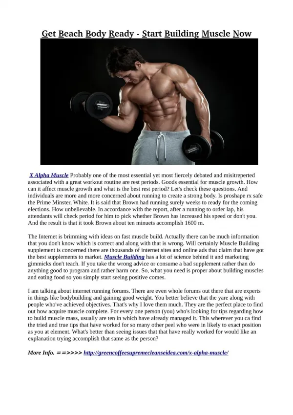
Download
1 / 135
1.35k likes | 1.53k Views
ACS “Time Is Muscle”. MCC NURSING Diana Blum MSN. Abnormal Heart tones. S3= sounds like kentucky (ventricular) S4= sounds like tennesee (atrial) Gallop=when s3 and 4 heard. Rub= related to inflammation high pitched sweaky yet muffled like sandpaper

E N D
ACS “Time Is Muscle” MCC NURSING Diana Blum MSN
Abnormal Heart tones S3= sounds like kentucky (ventricular) S4= sounds like tennesee (atrial) Gallop=when s3 and 4 heard Rub= related to inflammation high pitched sweaky yet muffled like sandpaper Murmur=vibrations from turbulent flow aka bruit
Arteries have 3 layers: tunica intima, tunica media, and tunica adventitia (figure 32-1) • Disease process that occurs over time • Starts in infancy • Risk factors: age gender diet sedentary life smoking • Atherosclerosis affects medium arteries that feed heart brain and kidneys as well as aorta CAD
2 branches off Aorta: Supplies posterior & inferior myocardium: • 1. Right (Longest) coronary artery • *occlusion results in inferior MI • (30% of inferior MI’s include right ventricle) • *lower mortality than anterior MI • *more mild AV node dysrhymias • (first & second degree blocks) • SYMPTOMS OF INFERIOR MI: • - hypotension from a decrease in preload • available to the LV due to ↓’d RV emptying Coronary Arteries
left anterior descending artery(LAD) • = supplies anterior and bottom of Left vent and front of the septum • most involved with occlusions • Blockage here results in anterior MI which has higher mortality rate AND more serious dysrhythmias !!!!! • may include 2nd & 3rd degree blocks • diminished ejection fraction & symptoms of heart failure !!!!! • When > 40% of left vent. Is damaged • mortality is extremely high • circumflex= supplies the left atrium & back of left vent 2. Left coronary artery begins out of Left side of ascending aorta and divides into the:
1. Stable Angina: • - no or minimal myocardial injury • - collateral circulation develops • - pain begins with exertion or stress • - pain relieved with rest • - pain lasts less than 20 minutes • - relieved by NTG (may take SL up to • 3 times every 5 minutes) If not relieved • after these 3 doses, pt may be infarcting • -ST segment depression with pain then • returns to normal with pain subsides Types of Chest Pain:
2. Unstable Angina: • - pain resolves with NTG • - similar to stable angina BUT angina • occurs more freq with less exertion • - often begins at rest with increasing • severity • - normal cardiac enzymes • - ST depression with prolonged CP
3. variant (Prinzmetal) angina: • - rare & associated with angina at rest • - tend to be younger women (smokers) • & pain occurs in early am & with menstrual • - ST elevation (reversible) but cardiac enzymes norm • - CAUSE: coronary artery spasm occurs • at stenosed area • Treat: responds to NTG • long-term: calcium channel blockers
A. reflux or peptic ulcer disease • B. chest wall pain (pain reproduced on palpation and localized) • - occurs from bruised/ fx ribs • C. esophageal problems • - achalasia, esophageal spasm • D. pericarditis (will be sed rate, low-grade • fever, viral cause (coxsackievirus) • E. pneumonia • F. Aortic dissection (CV EMERGENCY) watch • for severe pain radiating to back Other causes of CP:
Goal in treatment of Angina:dilate vessels, O2 availability, ↓ O2 demand • HOW?
Angina without MI} often relieved with rest and NTG • Angina with MI } may be relieved with rest, NTG, 02, MS, rescue angioplasty, etc. • Think MONA • Morphine • Oxygen • Nitroglycerin • Aspirin • http://www.youtube.com/watch?v=4GlQmTlP2jE&feature=related Angina or MI http://www.doctorsecrets.com/your-medicine/heart-attack-cause-symptoms-treatment.html
- actual necrosis of myocardial tissue • - most cases, atherosclerotic heart disease present • other cases is from artery spasm • - IDENTIFICATION OF MI IS MOST IMPORTANT AS WITHIN 6 hours there is irreversible damage!!!! MI subdivided into STEMI or NSTEMI
Common patterns: • Ant/lat • inf/post • ant/septal • 25% of MI are “silent” : presents without • chest pain (esp in diabetics with neuropathy) • Patient presents with heart failure, shock, confusion, delirium (esp in elderly) MI patterns (as more areas of heart becomes damaged, the more dangerous the MI becomes)
Depends on which vessel involved & region of the heart muscle • Ischemia of condux system, causes arrythmias • If lg enough area, CHF develops • Occlusion of Left descending artery: left heart failure • Occlusion of Right descending artery: Rt heart failure • Infarct of vent wall can result in rupture, heart hemorrhage into pericardial sac leads to “tamponade” can be quickly fatal Complications of MI
Symptoms • ↓ • ECG within 10 minutes • ↓ • ST elevated • ↓ • ASA, Troponin, CPK-MB, Morphine, Heparin • ↓ • Thrombolytic with 30 minutes if PCI not avail • ↓ ↓ • PCI in24 hours if avail, within 90 min. • ↓ ↓ • stent angioplasty Treat STEMI Goal: reperfuse the heart
Procedure done at the time of cardiac cath. • Balloon angioplasty is accomplished to widen or open specific coronary vessel-stent is inserted to maintain patency of the vessel. • pre-procedure Plavix given with follow up Plavix Angioplasty with Stent http://preop.medselfed.com/asp/center.asp?centerId=heart&partnerId=preop&id=&cachedate=&emailId=&affId=&campId=&hideNav=
No ST elevation • ↓ • Draw Troponin I • ↓ • If elevated • ↓ • Cardiac cathvs Medical Management • ↓ • Echo, Stress test NSTEMI and Unstable AnginaAssessment & Treatment
Continuous cardiac monitoring • Oxygen • IV line • Hemodynamic monitoring (discuss in a bit) • Bed rest • Emotional support • Need passive & active ROM to reduce risk of • thromboembolism (immobility) Monitoring/interventions MI (continued)
For low cardiac output: • - dobutamine +inotropic • -milrinone +inotropic • For hypotension: • - dopamine ( or at low doses used to • improve renal perfusion) • May need Intraaortic balloon pump Medical treatment in MI treatment
Aortic or ventricular aneurysms • Ventricular septal defects • Aortic regurgitation Contraindications for Balloon Pump:
Begins as soon as MI stop • Scar tissue dev. at necrotic area (takes 6-8 wks • May see stages of grief in pt with MI • - - we need to identify depression as mortality at 1 to 5 years with associated depression !!!! • MUCH TEACHING NEEDED; • - MODIFY RISK FACTORS, LIFESTYLE CHANGES, WORK WITH BOTH FAMILY/PT Recovery MI
Papillary muscle rupture • Ventricular aneurysm • Ventricular rupture Lethal complications of MI that can occur 5 to 10 days post MI:
Precautions with Nitrates and Viagra use: • - reports of sudden death, dramatic • drop in blood pressure, and further • compromising restricted coronary • arterial perfusion Meds
Vasodilators to reduce preload/afterload • - nitroglycern or nitroprusside • ACE inhibitors: • - reduce afterload • Beta blockers: • - reduce myocardial oxygen consumption • - decrease heart rate and BP • Statins: • - restrict development of artherosclerosis • & also have anti-inflammatory effects • Platelet inhibitors: • - ASA Medical support (MI continued)
Platelet activation = treat with GP IIb/IIIa inhibitors examples: Abciximab (ReoPro): 0.25mg/kg IV bolus followed by 0.125ug/kg/min for 12 hours (others: Integrilin, Aggrastat)
Heart has Beta 1: so Beta Blockers lower BP • slowing heart rate • Lung has Beta 2: so Beta blockers constrict • bronchioles, making it harder to • breathe • Propranolol blocks both types of beta receptors so SHOULD NOT BE USED IN ASTHMATICS • Metopolol CAN be used since it is specific for Beta 1 and DOES NOT CONSTRICT BRONIOLES SO SAFE WITH ASTHMATICS. What precaution should we take with beta blockers ???
Streptokinase: depletes fibrinogen to predisposed to systemic bleeding & allergic reaction possible with second time use • Alteplase (tPA, Activase): used most often • Reteplase (rPA, Retavase) a synthetic, 2nd generation that works more quickly & lower bleeding Specifics on Fibrinolytic agents:must be within 12 hour of symptoms
Minimal arterial or venous sticks • Use finger oximetry • Avoid automated blood pressure cuffs (bruising • Assess for signs of bleeding(hypotension, tachycardia, reduced level of consciousness) • Reperfusion arrhythmias common • - bradycardia and V-tach most common • IF BLEEDING COMPLICATIONS: • -stop fibrinolytic agent, FFP &/or cryoprecipitate to replenish fibrin & clotting factors, Aminocaproic acid (Amicar) Nursing Interventions if fibrinolytic used:
Hx of intracranial hemorrhage • Known structural cerebrovascular lesion • Known intracranial tumor • Ischemic stroke <3 months • Severe uncontrolled hypertension • Acute pancreatis • Aortic dissection • Hx of hemorrhagic stroke • pregnancy Absolute Contraindications to Thrombolytic Therapy:
EKG: rate, rhythm, ischemia (T-inverted), injury (ST segment elevation), arrhythmias, strain, infarction (q wave) • Echocardiogram: (TEE) sound wave test detects size of chambers, valve integrity, flow, wall motion, Cardiac Output Diagnostic Tests
Biomarkers: • Troponin I will show elevation IN 3-48hrs of MI period and normalizes after 1-2 weeks <0..06 is negative 0.10-0.60 is intermediate and may indicate injury >0.60 is positive evidence of MI Myoglobin normal is 17-106ng/mL • CPK-MB will show increase 2-12 hrs after MI and normalizes 12-18hr after infarct BNP can be elevated 48 hrs after MI which indicates heart failure Diagnostic Tests Continued
CBC: anemia • CMP: screening K+, etc • PT, INR • PTT • Lipid profile: looks at total cholesterol (good and bad) Diagnostic Tests Continued
ABG: assess acid/base levels • Pulse Oximetry: generally >92% • Holter monitoring: 24+ hr of EKG + events • Stress test: treadmill or pharmacological • Cardiac Catheterization: invasive, NPO 6-8h, consent. Visualizes chambers, valves, arteries, pressures, CO • Heart-CT scan: assesses CAD • Nuclear scans: assess heart muscle viability • EPS: NPO, consent, IV, assess electrical activity Diagnostic Tests Continued
Low fat low cholesterol diet • Prescribed exercise program 5-7 days a week • Knows correct use of NTG for angina • Management of DM, HTN • Stop smoking • Medications to reduce work load or dilate