Download
1 / 15
160 likes | 316 Views
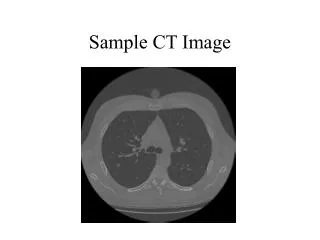
Enhanced CT Image advantages and potential. Reinhard Ruf Siemens AG Healthcare Sector Chair of Working Group 21. Enhanced CT Image. Introduction Use Cases (CT Cardio, Neuro Stroke) Conclusions. Enhanced CT Image. Introduction Supported Use Cases and Experiences Conclusions.

E N D
Enhanced CT Imageadvantages and potential Reinhard Ruf Siemens AGHealthcare Sector Chair of Working Group 21
Enhanced CT Image • Introduction • Use Cases (CT Cardio, Neuro Stroke) • Conclusions DICOM International Conference & Seminar
Enhanced CT Image • Introduction • Supported Use Casesand Experiences • Conclusions DICOM International Conference & Seminar
Enhanced CT Image • Introduction • Enhanced MR, CT, US, XA, XRF, PET • Efficient way of storing unified parameters together with image data • Support of complex use cases DICOM International Conference & Seminar
Enhanced CT Image • Introduction • Use Cases (CT Cardio, Neuro Stroke) • Conclusions DICOM International Conference & Seminar
Enhanced CT Image • Use CasesCardio CT • Saving of cardio result volumes like • Single volume • Multi phase study • Save cardiac and respiratory synchronization information. • Used to display heart beat animation DICOM International Conference & Seminar
Enhanced CT Image DICOM International Conference & Seminar
Enhanced CT Image • Cardiac synchronization information: DICOM International Conference & Seminar
Enhanced CT Image DICOM hint to how to use dimensions: • For example, the attribute specifying temporal position of frames may be any appropriate temporal attribute, such as Nominal Cardiac Trigger Delay Time (0020,9153) or Nominal Percentage of Cardiac Phase (0020,9241) in the Cardiac Synchronization Sequence (0018,9118) if the temporal position of frames is referenced to the cardiac R-wave, or Nominal Respiratory Trigger Delay Time (0020,9255) or Nominal Percentage of Respiratory Phase (0020,9245) in the Respiratory Synchronization Sequence (0020,9253) if the temporal position of frames is referenced to the latest inspiration maximum. DICOM International Conference & Seminar
Enhanced CT Image • Use CasesNeuro Stroke • Saving of perfusion result volumes like • CBF(Cerebral Blood Flow) • CBV(Cerebral Blood Volume) • TTP(Time to Peak) etc. • Save color LUT informationtogether with unit information. • LUT is applied, the displayed images can show appropriate units. DICOM International Conference & Seminar
Enhanced CT Image DICOM International Conference & Seminar
Enhanced CT Image DICOM hint to use presentation state objects: • In order to annotate images, whether during acquisition or subsequently, SOP Instances of the Grayscale Softcopy Presentation State Storage or the Structured Report Storage SOP Classes that reference the image SOP Instance, may be used. No standard mechanism is provided for inclusion of annotations within the image SOP Instance itself, and implementers are discouraged from using private extensions to circumvent this restriction. Grayscale Softcopy Presentation State Storage Instances that are generated during acquisition may be referenced from the Image SOP Instance by using the Referenced Grayscale Presentation State Sequence in the Enhanced CT Image Module. See C.8.15.2. DICOM International Conference & Seminar
Enhanced CT Image • Introduction • Use Cases (CT Cardio, Neuro Stroke) • Conclusions DICOM International Conference & Seminar
Enhanced CT Image • Conclusions • Improve interoperability (color, real world value mapping, synchronization technique, etc.) • Support of time / respiratory synchronized volume representation • Grayscale images with color information • Combined functionality with Presentation State Objects (e.g. graphics, annotations, blending, …) • Even more potential and use cases together with Multi-Dimensional Presentation State Object (WG 11, Supplement 156) DICOM International Conference & Seminar
References http://medical.NEMA.org/ http://www.HL7.org/ http://www.IHE.net/ Thank you for your attention ! DICOM International Conference & Seminar