Download
1 / 74
740 likes | 888 Views
Cardiovascular Emergencies Part 1. Assessment. Primary survey/ resuscitation Secondary survey. Factors For Consideration. Unmodifiable Age Sex Heredity Race. Modifiable B/P Obesity Dyslipidemia Smoking Sedentary life style Stress Diabetes. Focused Survey. Subjective data

E N D
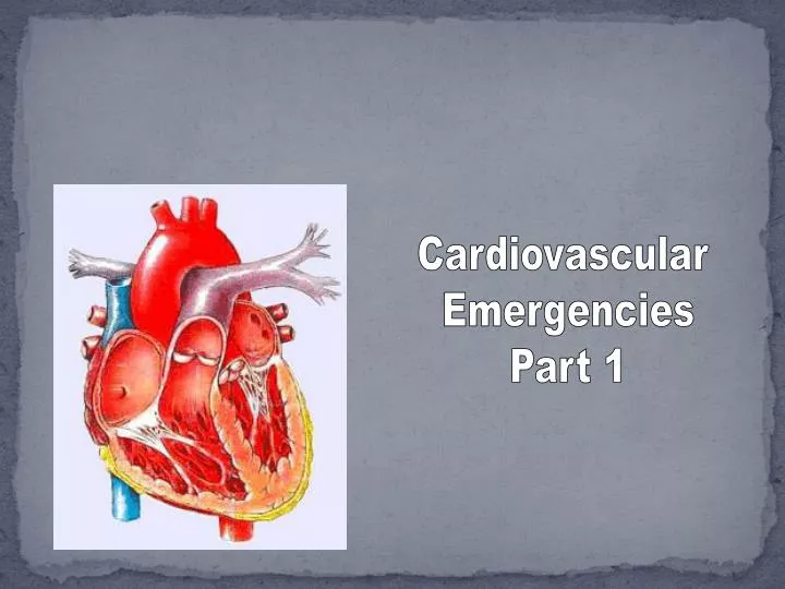
Cardiovascular Emergencies Part 1
Assessment • Primary survey/ • resuscitation • Secondary survey
Factors For Consideration • Unmodifiable • Age • Sex • Heredity • Race • Modifiable • B/P • Obesity • Dyslipidemia • Smoking • Sedentary life style • Stress • Diabetes
Focused Survey Subjective data • Chief complaint • History of present illness • Onset: OPQRST • Pain: PQRST • Provocation • Quality • Region/radiation • Severity • Time • Duration • Development over time • Periodicity (? Comes/goes)
Focused Survey (Continued) • Dyspnea • SOB • Dyspnea on exertion • Positional dyspnea • Paroxysmal nocturnal dyspnea • Orthopnea • Cough • Dry, “cardiac” cough • Hemoptysis • Syncope • Palpitations • Fatigue Nausea and Vomiting • Headache • Behavioral change • Activity limitations • Injury: mechanism and time
Medical History • Coronary Heart Disease • Angina • Previous MI • Hypertension • CHF • Pulmonary Disease • Diabetes • Renal Disease • Previous Cardiac Surgery • Congenital Anomalies • Allergies
Current/Past Medication Use • Nitrates • Beta Blockers • Calcium Channel Blockers • Anti- hypertensives • Digitalis • Diuretics • Antidysrhythmics • Anticoagulants • Steroids • Specific Pulmonary Drugs • Illicit Drugs • OVC Medications
Physical Exam • General Survey • LOC • Respiratory status • Rate, regularity, effort, breath sounds • Skin • Color, temperature, moisture, capillary refill • Edema • Dependent, extremities, sacrum, pleural effusion, ascitis, cardiac (pitting)
Physical Exam 0 = absent 1 = thready 2 = normal 3 = bounding • Cyanosis • Central • Peripheral • Clubbing • B/P Measurement • Both arms • Orthostatic (supine, sitting, standing)
Physical Exam • Apical heart rate • Regular, irregular, regularly irregular, irregular irregular • Peripheral pulses • Pupils • Size, equality, reactive
Inspection • Tracheal position • Neck veins • Thorax • Configuration • Deformities, anterior, posterior, A-P diameter, symmetrical movement • Injuries, penetrating, blunt (ecchymosis, contusions) evidence of scars, surgery • Abnormal chest movement, asymmetrical, paradoxical
Physical Exam (Inspection) • Precordium • Apical pulse • Abnormal precordial movements, heaves, lifts, pulsations, retractions • Epigastrium, pulsations
Physical Exam: Palpation • Areas tender, or crepitus • Epigastrium
Physical Exam: Auscultation • Auscultatory sites • Murmurs: systolic/diastolic • Variations in rhythm • Extra sounds • Pericardial friction rub • Venous hum • Arterial bruits • Clicks
Physical Exam: Diagnostic Procedures • ECG, 12 lead • Rate, rhythm • Presence of cardiac dysrhythmias • Evidence of myocardial ischemia, injury • Presence of intraventricular conduction defect • Evidence of previous MI
Physical Exam:Diagnostic Procedures • CBC • Serum electrolytes • Cardiac serum markers • Troponin • Basic metabolic profile • Coagulation studies • Digoxin level • Serum creatinine, BUN • T& C • ABGs • Routine urinalysis
Diagnostic Procedures • Pulse oximetry • Radiography • Chest x-ray • Heart size and location • Presence of edema • Pulmonary infiltrates • Pleural effusions • Air and fluid levels in trauma patients • Mediastinal width • Bony structure integrity • Cardiac catheterization • Echocardiogram
Interventions • Determine priorities • Airway, vital signs, cardiac rhythm, ABGs, Pulse oximetry, • Control pain • Relieve anxiety • Education patient/other • Prevent complications • Establish care plan • Emergency equipment • Initiate appropriate interventions • Document data • Monitor responses and adjust
Pediatrics • Growth & Development • R/T congenital heart disease (heart defects) • Acquired heart disease (rheumatic fever) • Endocrine (diabetes) • Other • Drug ingestions, ex: tricyclics, digoxin • Trauma (falls, MVCs) • Suffocation (plastic bags, drowning, accidental hanging)
Pediatric • Pearls: • Cardiac arrest usually d/t progressive deterioration in respiratory and heart function • CHF, cardiogenic shock, dysrhythmias are unusual. If occur congenital. • Immature conduction system and autonomic innervation may contribute to dysrhythmias
Geriatric • Presence of chronic diseases • Altered drug metabolism • Multiple physiological differences and changes in lab values must be considered when assessing the older patient • Psychological and social changes: patient may have different goals for their treatment, discuss with patient • Geriatric patients can adapt to disease so well that symptoms are not obvious • Arteriosclerotic changes in aorta and peripheral pulses may pose a difficulty in palpating • Rhythm abnormalities are so common that they may be “normal”
Pearls • “Go slow, stay low” with medications • Concurrent use of other medications cause problems, • Easy to use meds are helpful (transdermals) • One time or two time doses daily • Evaluate medications on a frequent basis
Angina • Stable • Symptom of ischemia • “pain or discomfort” • Poorly localized • Flow/Demand imbalance • May be chronic, acute, or unstable
Angina (continued) • Unstable angina • New symptoms of angina • Increasing symptoms that occur at rest or with on exertion • Usually due to platelet aggregation • Leads to atypical chest pain
Angina (Continued) • Unstable angina diagnosis • Angina is at rest, as well as minimal exertion (usually 20 minutes or longer) • Angina of new onset (several weeks), starting with physical exertion, and markedly limits activity • Previously diagnosed stable angina
Angina cont’d • Variant Angina • May or may not be due to atherotic changes • Thought to be due to coronary spasm • Prinzmetal’sangina • May occur at the same time, daily • Usually not associated with exertion or stress • Occurs at younger ages • ST elevation seen during pain, then disappears
Angina: Curveballs • GERDs • Biliary Colic • Chest Wall Pain • Pericarditis • PE • Aortic Dissection • Dysrhythmias
Diagnostic Procedures: ECG • ST depression may accompany pain with stable angina • Transient ST-segment deviations (depression or elevation), and T wave inversion occur commonly with unstable angina • Variant angina: ST elevation occurs with pain, subsides when pain does • May see LV hypertrophy, old MI, nonspecific ST and T-wave abnormalities and AV defects
Diagnostic Procedures (Continued) • CBC • Cardiac serum markers…no elevation should occur unless cell damage • Chest X-ray ( CHF, cardiomegaly)
Interventions • Continuous monitoring • O2 • IV, Draw labs • 12 Lead ECG • Rest • Decrease anxiety • SL NTG, B/P 100 mm HG followed by a drip • Assess for H/A • Reflex tachycardia • Cautious with elderly
Interventions (continued) • Beta blockers • If clinical situation deteriorates after B Blocker, consider coronary artery spasm • Assess for signs of heart failure • Adverse effects of blockers are considered more common and severe in geriatric
Interventions (continued) • Administer antiplatelet agents • ASA (4-5 baby aspirin) • Administer ASAP • Decreases platelet activation and thrombus formation • TEACH/EDUCATION
MI: Data • Physical exam • General appearance: Anxious, restless, clenched fist against chest (Levine sign) Look of doom • Heart rate: may be ok, tachycardia (most common), bradycardia (inferior and RV), Regular or irregular PVCs common
Data (continued) • Arterial BP • Majority of patients with uncomplicated MI are normotensive • May be elevated due to SNS stimulation • Pain and anxiety • Decreased as a result of impaired cardiac function or due to drug administration (nitrates, M.S.)
Data (continued) • Respiratory rate: Initially elevated. Should return to normal after pain relief • Patients with CHF, respiratory rate correlates with severity of condition
Data (continued) • Peripheral: How bad is the patient’s condition? • Pallor, cyanosis, diaphoreses, mottled, cool, peripheral pulses variable • Temperature: Often increases 4-8 hours post MI • Heart Sounds: muffled. murmurs may be transient or permanent
Diagnosis • Continuous cardiac monitoring • 12 lead ECG • Determine Location of infarct (next slide)
Serum Markers • Myoglobin • Elevated 1-4 hours after onset of MI, peaks 6-7 hours, normal in about 1 day • Lacks specificity, found in skeletal muscle
Serum Markers (continued) • Troponins: • Most recent marker, most sensitive and specific for cardiac damage • Elevated 3-12 hours after MI, Peaks in 24 hours, with TnI , returns to normal in 5-12 days • TnT may be elevated in patients with renal failure, which is not the case with TnI; therefore TnI is utilized.