Download
1 / 59
600 likes | 834 Views
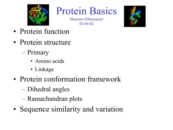
Protein Basics. Protein function Protein structure Primary Amino acids Linkage Protein conformation framework Dihedral angles Ramachandran plots Sequence similarity and variation. Protein Function in Cell. Enzymes Catalyze biological reactions Structural role Cell wall

E N D
Protein Basics • Protein function • Protein structure • Primary • Amino acids • Linkage • Protein conformation framework • Dihedral angles • Ramachandran plots • Sequence similarity and variation
Protein Function in Cell • Enzymes • Catalyze biological reactions • Structural role • Cell wall • Cell membrane • Cytoplasm
Hemoglobin – Quaternary Structure Two alpha subunits and two beta subunits (141 AA per alpha, 146 AA per beta)
Hemoglobin – Tertiary Structure One beta subunit (8 alpha helices)
Hemoglobin – Secondary Structure alpha helix
Hemoglobin – Primary Structure NH2-Val-His-Leu-Thr-Pro-Glu-Glu- Lys-Ser-Ala-Val-Thr-Ala-Leu-Trp- Gly-Lys-Val-Asn-Val-Asp-Glu-Val- Gly-Gly-Glu-….. beta subunit amino acid sequence
Protein Structure - Primary • Protein: chain of amino acids joined by peptide bonds
Protein Structure - Primary • Protein: chain of amino acids joined by peptide bonds • Amino Acid • Central carbon (Cα) attached to: • Hydrogen (H) • Amino group (-NH2) • Carboxyl group (-COOH) • Side chain (R)
General Amino Acid Structure H H2N COOH Cα R
Amino Acids • Chiral
Chirality: Glyceraldehyde D-glyderaldehyde L-glyderaldehyde
Amino Acids • Chiral • 20 naturally occuring; distinguishing side chain
Amino Acids • Chiral • 20 naturally occuring; distinguishing side chain • Classification: • Non-polar (hydrophobic) • Charged polar • Uncharged polar
Peptide Bond • Joins amino acids
Peptide Bond • Joins amino acids • 40% double bond character • Caused by resonance
Peptide bond • Joins amino acids • 40% double bond character • Caused by resonance • Results in shorter bond length
Peptide bond • Joins amino acids • 40% double bond character • Caused by resonance • Results in shorter bond length • Double bond disallows rotation
Protein Conformation Framework • Bond rotation determines protein folding, 3D structure
Protein Conformation Framework • Bond rotation determines protein folding, 3D structure • Torsion angle (dihedral angle) τ • Measures orientation of four linked atoms in a molecule: A, B, C, D
Protein Conformation Framework • Bond rotation determines protein folding, 3D structure • Torsion angle (dihedral angle) τ • Measures orientation of four linked atoms in a molecule: A, B, C, D • τABCD defined as the angle between the normal to the plane of atoms A-B-C and normal to the plane of atoms B-C-D
Protein Conformation Framework • Bond rotation determines protein folding, 3D structure • Torsion angle (dihedral angle) τ • Measures orientation of four linked atoms in a molecule: A, B, C, D • τABCD defined as the angle between the normal to the plane of atoms A-B-C and normal to the plane of atoms B-C-D • Three repeating torsion angles along protein backbone: ω, φ, ψ
Backbone Torsion Angles • Dihedral angle ω : rotation about the peptide bond, namely Cα1-{C-N}- Cα2
Backbone Torsion Angles • Dihedral angle ω : rotation about the peptide bond, namely Cα1-{C-N}- Cα2 • Dihedral angle φ : rotation about the bond between N and Cα
Backbone Torsion Angles • Dihedral angle ω : rotation about the peptide bond, namely Cα1-{C-N}- Cα2 • Dihedral angle φ : rotation about the bond between N and Cα • Dihedral angle ψ : rotation about the bond between Cα and the carbonyl carbon
Backbone Torsion Angles • ω angle tends to be planar (0º - cis, or 180 º - trans) due to delocalization of carbonyl pi electrons and nitrogen lone pair
Backbone Torsion Angles • ω angle tends to be planar (0º - cis, or 180 º - trans) due to delocalization of carbonyl pi electrons and nitrogen lone pair • φ and ψ are flexible, therefore rotation occurs here
Backbone Torsion Angles • ω angle tends to be planar (0º - cis, or 180 º - trans) due to delocalization of carbonyl pi electrons and nitrogen lone pair • φ and ψ are flexible, therefore rotation occurs here • However, φ and ψ of a given amino acid residue are limited due to steric hindrance • Only 10% of the area of the {φ, ψ} space is generally observed for proteins • First noticed by G.N. Ramachandran
G.N. Ramachandran • Used computer models of small polypeptides to systematically vary φ and ψ with the objective of finding stable conformations • For each conformation, the structure was examined for close contacts between atoms • Atoms were treated as hard spheres with dimensions corresponding to their van der Waals radii • Therefore, φ and ψ angles which cause spheres to collide correspond to sterically disallowed conformations of the polypeptide backbone
Ramachandran Plot • Plot of φ vs. ψ • Repeating values of φ and ψ along the chain result in regular structure • For example, repeating values of φ ~ -57° and ψ ~ -47° give a right-handed helical fold (the alpha-helix) • The structure of cytochrome C-256 shows many segments of helix and the Ramachandran plot shows a tight grouping of φ, ψ angles near -50, -50
The structure of cytochrome C-256 shows many segments of helix and the Ramachandran plot shows a tight grouping of φ, ψ angles near -50,-50 cytochrome C-256 Ramachandran plot alpha-helix
Ramachandran Plot • White = sterically disallowed conformations (atoms in the polypeptide come closer than the sum of their van der Waals radii) • Red = sterically allowed regions (namely right-handed alpha helix and beta sheet) • Yellow = sterically allowed if shorter radii are used (i.e. atoms allowed closer together; brings out left-handed helix)