Download
1 / 8
90 likes | 329 Views
Lab #2. Bacteriology & the Archaea. PROTEOBACTERIA. Bacterial Groups. Subgroup: Alpha Proteobacteria. 2.5 µm. Rhizobium (arrows). 1. Proteobacteria: diverse group of gram negative bacteria a. alpha: live in close associated with eukaryotes

E N D
Lab #2 Bacteriology & the Archaea
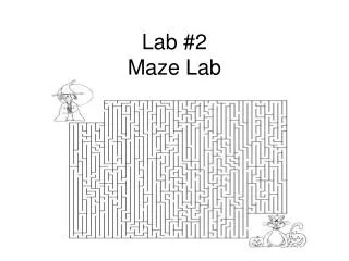
PROTEOBACTERIA Bacterial Groups Subgroup: Alpha Proteobacteria 2.5 µm Rhizobium (arrows) • 1. Proteobacteria: diverse group of gram negative bacteria • a. alpha: live in close associated with eukaryotes • Rhizobium – lives in nodules within the roots of legumes – convert atmospheric N2 into compounds that the plants can use (nitrogen fixation) • some strains can cause tumors in plants – Agrobacterium – used to genetically modify plants • b. beta: nutritionally diverse • Nitrosomas – soil bacteria that plays a role in N2 recycling by oxidizing NH4 into NO2- • c. delta: slime secreting myxobacteria • when the soil dries out – they form into aggregations called fruiting bodies – release spores into the environment • establishment of new colonies in better environments • bdellovirbio bacteria “charge” at other bacteria at speeds equivalent to 240km/hr • drills into its prey using its flagella and digestive enzymes Subgroup: Beta Proteobacteria 1 µm Nitrosomonas Subgroup: Gamma Proteobacteria 0.5 µm Chromatium Subgroup: Delta Proteobacteria 5 µm 10 µm Bdellovibrio bacteriophorus Chrondromyces crocatus Subgroup: Epsilon Proteobacteria 2 µm Heliocobacter pylori
PROTEOBACTERIA Subgroup: Alpha Proteobacteria 2.5 µm Rhizobium (arrows) • 1. Proteobacteria: diverse group of gram negative bacteria • d. gamma: autotrophic & hetertrophic species • include the older classification known as sulfur bacteria (e.g. Thiomargarita namibiensis) • these obtain energy by oxidizing H2S – producing sulfur as a waste • many heterotrophic strains are pathenogenic (e.g. Legionella, Salmonella and Vibrio cholerae) • non pathenogenic strain = E. coli • e. epsilon: many are pathenogenic to humans and other animals • includes Campylobacter = blood poisoning • Helicobacter pylori = stomach ulcers • f. zeta: relatively new classification Subgroup: Beta Proteobacteria 1 µm Nitrosomonas Subgroup: Gamma Proteobacteria 0.5 µm Chromatium Subgroup: Delta Proteobacteria 5 µm 10 µm Bdellovibrio bacteriophorus Chrondromyces crocatus Subgroup: Epsilon Proteobacteria 2 µm Heliocobacter pylori
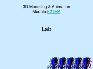
Bacterial Groups CHLAMYDIAS 2.5 µm Chlamydia (arrows) SPIROCHETES • 2. Gram positive bacteria: rival proteobacteria in diversity • 5 major subgroups • two strains of Actinomycetes cause leprosy and tuberculosis – most decompose organic matter in soil • Streptomyces used by pharmaceutical companies to produce antibiotics • numerous strains are very pathogenic: Bacillus anthracis, Clostridium botulinum, Staphylococcus and Streptococcus • 3. Chlamydias • can only survive in animal cells – depend on their hosts for ATP • Chlamydia trachomatis – cause of nongonococcal urethritis (most common STD) • 4. Spirochetes • move through rotation provided by internal flagella-like filaments • Treponema pallidum – causes syphillis • Borrelia burgdorferi – causes Lyme disease • 5. Cyanobacteria • photoautotrophs • only prokaryotes with plant-like, oxygen-generating photosynthesis • abundant components of fresh water and marine phytoplankton 5 µm Leptospira GRAM-POSITIVE BACTERIA CYANOBACTERIA 50 µm Oscillatoria 5 µm Mycoplasmas covering a human fibroblast cell Streptomyces
Bacterial classification • colony morphology • bacterial colonies grow from single cells • colony is composed of millions of bacteria • each colony has a characteristic size, sheep, consistency, texture and color • common colony shapes: • punctiform = each colony is less than 1mm • round • filamentous – often confused with fungus (which is more “fuzzy”) • irregular
Bacterial classification • cell morphology • bacilli (rod) • cocci (spherical) • spirilla • many cells adhere to each other and form clusters or chains • under some environments – many different species may associate with each other – creating a community called a biofilm • biofilms are usually found where nutrients are plentiful • soils, water pipes, surface of your teeth
Gram staining 1. Place a slide with a bacterial smear on a staining rack. 2. STAIN the slide with crystal violet for 1-2 min. 3. Pour off the stain and rinse with water thoroughly.4. Flood slide with Gram's iodine for 1-2 min. 5. Pour off the iodine and rinse with water thoroughly.. 6. Decolourize by washing the slide briefly with acetone (2-3 seconds) – alternatively use 95% ethanol 7. Wash slide thoroughly with water to remove the acetone 8. Flood slide with safranin counterstain for 2 min. 9. Wash with water. 10. Blot excess water and dry by hand over bunsen flame. • both Gram-positive and Gram-negative bacteria take up the same amounts of crystal violet (CV) and iodine (I). • CV-I complex is trapped inside the Gram-positive cell by the washing of the bacteria with 95% ethanol – results in the dehydration and reduced porosity of the thick cell wall – limits the loss of CV-I complex – PURPLE STAIN • thin peptidoglycan layer of the gram negative bacteria does not impede extraction of the CV-I complex • plus the outer membrane limits the amount of CV-I complex that can reach the PG layer – CLEAR STAIN http://www.youtube.com/watch?v=OQ6C-gj_UHM