Download
1 / 48
530 likes | 882 Views
052. Epithelium and glands. Dr. Larry Johnson. Objectives. Identify every epithelium present in any tissue section. Differentiate between mucus and serous secreting epithelia. Identify single-celled glands, endocrine glands and the various types of exocrine glands.

E N D
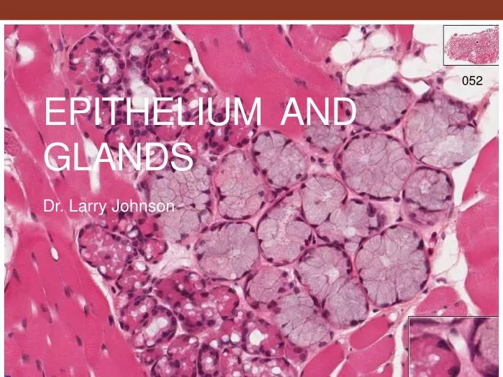
052 Epithelium and glands Dr. Larry Johnson
Objectives • Identify every epithelium present in any tissue section. • Differentiate between mucus and serous secreting epithelia. • Identify single-celled glands, endocrine glands and the various types of exocrine glands. • Detail the structure of the sebaceous gland. • Identify the different types of sweat glands and distinguish the duct from the secretory region. • Identify myoepithelial cells and know their function. From: Douglas P. Dohrman and TAMHSC Faculty 2012 Structure and Function of Human Organ Systems, Histology Laboratory Manual
ORIGIN AND DISTRIBUTION OF EPITHELIUM ECTODERM - EPIDERMIS OF SKIN AND EPITHELIUM OF CORNEA TOGETHER COVERS THE ENTIRE SURFACE OF THE BODY; SEBACEOUS AND MAMMARY GLANDS ENDODERM - ALIMENTARY TRACT, LIVER, PANCREAS, GASTRIC GLANDS, INTESTINAL GLANDS • ENDOCRINE GLANDS - LOSE CONNECTION WITH SURFACE MESODERM • ENDOTHELIUM - LINING OF BLOOD VESSELS • MESOTHELIUM - LINING SEROUS CAVITIES ECTODERM MESODERM ENDODERM
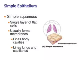
Characteristics of epithelium • Classification by # of layers • Simple = single layer • Stratified = multiple, stacked layers • Pseudostratified = appears to be multiple layers, but all cells touch basement membrane (all cells do not necessarily reach lumen) • Classification by shape • Squamous = flat • Cuboidal = square • Columnar = column
Laboratory Experience • Identify and characterize various types of epithelia • Recognize various glands and modes of secretion • Recognize gland associated structures/cells and differentiate glands from their ducts • Understand significance of cytological expression of epithelial cells with regard to function 053
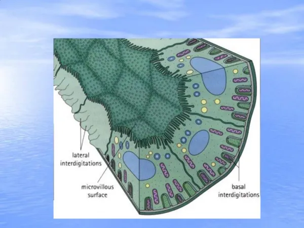
Slide 33: Kidney (PAS/hematoxylin) Periodic acid – Schiff • Used to visualize: glycogen, glycoproteins, and glycosaminoglycans • Steps in PAS staining • Periodic acid oxidizes 1,2-glycols to aldehydes • Schiff’s reagent colors the aldehyde groups (pink to magenta) Brush border • The brush border is composed of numerous microvilli which function to increase surface area for absorption of nutrient material and fluid. Basement membrane The structure on the brush border that stains with periodic-acid Schiff (PAS) stain is the glycocalyx.
EM 1 & 7 033 glycocalyx PAS staining Observe various brush border - microvilli • The brush border is composed of numerous microvilli of a uniform size projecting into a lumen. Each microvillus is composed of microfilaments (actin) surrounded by the cell membrane.
EM 2 Stereocilia of the epididymis - long thin microvilli • Stereocilia are very long, branched, non-motile microvilli of differing lengthsthat function in absorption from the lumen.
EM 5 & 6 Observe cell junctions characteristic of epithelium
SPECIALIZATION OF EPITHELIA MAINTAIN EXTENSIVE CONTACTS AMONG CELLS STRUCTURALLY AND FUNCTIONALLY POLARIZED JUNCTIONS ZONULA OCCLUDENS - TIGHT JUNCTION (BELT) ZONULA ADHERENS – ADHERING BELT DESMOSOME (MACULA ADHERENS) - SPOT ATTACHMENT GAP JUNCTIONS - COMMUNICATION
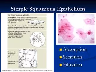
Slide 32: Kidney (H&E) Simple squamous Simple squamous Simple cuboidal Simple cuboidal
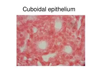
Slide 75: Thyroid gland (endocrine) Simple cuboidal epithelium Basement membrane
SUMMARY OF TISSUE FEATURES OF EPITHELIUM • AVASCULAR • EXTRANEOUS CELLS • REGENERATION • MIGRATION • METAPLASIA • BASEMENT MEMBRANE
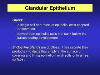
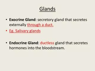
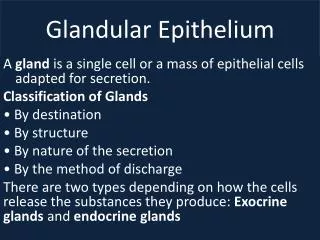
SECRETION – ACTIVE PROCESS CONSUMING ENERGY EXOCRINE GLANDS - DELIVER THEIR SECRETION INTO DUCTS OPENING INTO EXTERNAL OR INTERNAL SURFACE ENDOCRINE GLANDS - DUCTLESS, DELIVER THEIR SECRETIONS INTO THE LYMPH OR BLOOD STREAM PANCREAS has exocrine PANCREAS has endocrine
EXOCRINE GLANDS DUCT • SIMPLE - UNBRANCHED DUCT • COMPOUND - BRANCHED DUCT SECRETORY PORTION • TUBULAR • COILED TUBULAR • BRANCHED TUBULAR • ALVEOLAR • BRANCHED ACINAR • TUBULOACINAR • TUBULOALVEOLAR MUCUS VS SEROUS
TUBULAR • COILED TUBULAR • BRANCHED TUBULAR • ALVEOLAR • BRANCHED ACINAR • TUBULOACINAR • TUBULOALVEOLAR
ACINUS = FUNCTIONAL UNIT • SEROUS • MUCOUS 072 Mucous- Light staining Cytoplasm and dark, flattened nucleus at base of cell 072 Serous – dark red staining cytoplasm and lighter, spherical nucleus
Slide 72: Submandibular gland Stratified columnar epithelia Simple columnar epithelia
Slide 72: Submandibular gland Serous demilune Serous cells Mucous cells The submandibular gland is a mixed (seromucous) gland whose mode of secretion is merocrinesecretion =exocytosis without loss of cellular components
MECHANISM FOR RELEASE OF SECRETORY PRODUCTS MEROCRINE SECRETION – EXOCYTOSIS W/O LOSS OF SURFACE MEMBRANE APOCRINE SECRETION – LOSS OF PART OF APICAL CYTOPLASM AND SOME PLASMA MEMBRANE HOLOCRINE SECRETION – RELEASE OF WHOLE cell CYTOCRINE SECRETION – MELANIN GRANULES TRANSFERRED FROM MELANOCYTE TO KERATINOCYTES
APOCRINE MEROCRINE
CYTOCRINE SECRETION- PASS MELANIN GRANULES FROM MELANOCYTES TO KERATINOCYTES
Slide 61: Terminal Ileum Brush border composed of microvilli • Microvilli are fingerlike projections that greatly increase the surface area of certain cells to help increase absorption. Microvilli are non-motile and are composed of a core of thin microfilaments called actin. Goblet cell releasing contents Simple columnar epithelium
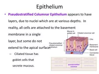
Slide 40: Trachea Goblet cell releasing contents Ciliated pseudostratified columnar epithelium with goblet cells • The main function of cilia is to sweep or move fluids, cells, or particulate matter across cell surface in the lumen as to remove dust in the lungs. Microtubules of the axoneme are at the core of cilia and make them motile. • . Note the thick basement membrane of the respiratory pseudostratified columnar epithelium.
Slide 93: Epididymis Pseudostratified columnar epithelium with stereocilia Microvilli: small, non-motile projections composed of thin microfilaments Stereocilia: long, non-motile projections (branched microvilli) composed of thin microfilaments Cilia: larger, long, motile projections composed of thick microtubules
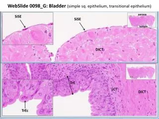
Slide 35: Urinary bladder Transitional epithelium Specialized “dome-shaped” cells Basal cells
Slide 34: Ureter Transitional epithelium
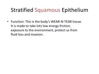
Stratified squamous epithelium: the protection epithelium • Keratinized stratified squamous (thick or thin) • Prevent dessication • Protect against abrasion • Prevent foreign invasion • Ex. Slide 29: Thick skin (ventral surface of finger) • Non-keratinized stratified squamous • Moist lubricated surface • Ex. Slide 52: Tongue
Slide 52: Tongue Non-keratinized stratified squamous epithelium Mucous acini Serous acini Mucous cells stain light and serous cells stain dark.
Slide 53: Esophagus Non-keratinized stratified squamous epithelium
Slide 29: Thick skin (ventral surface of finger) Epidermis with keratinized stratified squamous epithelium Dermis Hypodermis The major function of this type of epithelium (thick skin) is for protection from mechanical stress, but it also prevents dehydration.
Slide 29: Thick skin (ventral surface of finger) Keratinized stratified squamous epithelium Polyhedral cells Cuboidal cells of the stratum basale The cells in the stratum basale serve as stem cells for the epidermis, and their progeny differentiate as they move away from the base.
Slide 29: Thick skin (ventral surface of finger) Duct of eccrine sweat gland with stratified cuboidal epithelium Eccrine sweat gland Myoepithelial cells • Myoepithelial cells are eosinophilic because of the presence of a high density of contractile protein. These cells surround the gland like a net and expel glandular secretions upon contraction.
Slide 31: Thin skin (scalp) Sebaceous glands The mode of secretion used by sebaceous glands is holocrinesecretion Keratinized, stratified squamous epithelium
Slide 66: Recto-anal junction Anus – stratified squamous Rectum - simple columnar epithelium
Slide 66: Recto-anal junction Sebaceous glands Eccrine sweat gland Apocrine sweat gland
GLANDS OF EPIDERMAL ORIGIN SWEAT GLANDS • ECCRINE - COMMON SWEAT GLAND - LOCAL COOLING • APOCRINE AXILLARY REGION - FUNCTION IN ANIMALS, discharge in hair follicle
Epithelial tissues of the body • Use table as guide • Generalizations • Entire GI system from gastro-esophageal junction to recto-anal junction is lined by simple columnar epithelium • Cilia is present in most respiratory passages
EPITHELIA ARE SPECIALIZED FOR FUNCTIONS ABSORPTION - INTESTINE SECRETION - PANCREAS TRANSPORT - EYE, ENDOTHELIUM IN VESSELS EXCRETION - KIDNEY PROTECTION – AGAINST MECHANICAL DAMAGE AND DEHYDRATION SENSORY RECEPTION – PAIN TO AVOID INJURY, TASTE BUDS, OLFACTORY, ETC. CONTRACTION – MYOEPITHELIUM
SURFACE SPECIALIZATIONS OF EPITHELIA MICROVILLI - INTESTINE ABSORPTIVE CELL CILIA - RESPIRATORY EPITHELIUM BASAL LAMINA – ALL EPITHELIUM INTERCELLULAR CANALICULUS – HEPATOCYTE SECRETORY CANALICULUS – GASTRIC PARIETAL CELL FLAGELLA 038b
SURFACE SPECIALIZATIONS OF EPITHELIA INTERCELLULAR CANALICULUS – HEPATOCYTE
SURFACE SPECIALIZATIONS OF EPITHELIA SECRETORY CANALICULUS – GASTRIC PARIETAL CELL
Clinical Correlation Edward C. Klatt, M.D. Mercer University School of Medicine Normal larynx with ciliated pseudostratified columnar epithelium Abnormal larynx with stratified squamous epithelium
Many illustrations in these VIBS Histology YouTube videos were modified from the following books and sources: Many thanks to original sources! • Bruce Alberts, et al. 1983. Molecular Biology of the Cell. Garland Publishing, Inc., New York, NY. • Bruce Alberts, et al. 1994. Molecular Biology of the Cell. Garland Publishing, Inc., New York, NY. • William J. Banks, 1981. Applied Veterinary Histology. Williams and Wilkins, Los Angeles, CA. • Hans Elias, et al. 1978. Histology and Human Microanatomy. John Wiley and Sons, New York, NY. • Don W. Fawcett. 1986. Bloom and Fawcett. A textbook of histology. W. B. Saunders Company, Philadelphia, PA. • Don W. Fawcett. 1994. Bloom and Fawcett. A textbook of histology. Chapman and Hall, New York, NY. • Arthur W. Ham and David H. Cormack. 1979. Histology. J. S. Lippincott Company, Philadelphia, PA. • Luis C. Junqueira, et al. 1983. Basic Histology. Lange Medical Publications, Los Altos, CA. • L. Carlos Junqueira, et al. 1995. Basic Histology. Appleton and Lange, Norwalk, CT. • L.L. Langley, et al. 1974. Dynamic Anatomy and Physiology. McGraw-Hill Book Company, New York, NY. • W.W. Tuttle and Byron A. Schottelius. 1969. Textbook of Physiology. The C. V. Mosby Company, St. Louis, MO. • Leon Weiss. 1977. Histology Cell and Tissue Biology. Elsevier Biomedical, New York, NY. • Leon Weiss and Roy O. Greep. 1977. Histology. McGraw-Hill Book Company, New York, NY. • Nature (http://www.nature.com), Vol. 414:88,2001. • A.L. Mescher 2013 Junqueira’s Basis Histology text and atlas, 13th ed. McGraw • Douglas P. Dohrman and TAMHSC Faculty 2012 Structure and Function of Human Organ Systems, Histology Laboratory Manual - Slide selections were largely based on this manual for first year medical students at TAMHSC