Download
1 / 17
170 likes | 241 Views
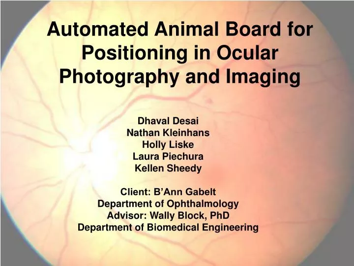
Automated Animal Board for Positioning in Ocular Photography and Imaging. Dhaval Desai Nathan Kleinhans Holly Liske Laura Piechura Kellen Sheedy Client: B’Ann Gabelt Department of Ophthalmology Advisor: Wally Block, PhD Department of Biomedical Engineering. Glaucoma.

E N D
Automated Animal Board for Positioning in Ocular Photography and Imaging Dhaval Desai Nathan Kleinhans Holly Liske Laura Piechura Kellen Sheedy Client: B’Ann Gabelt Department of Ophthalmology Advisor: Wally Block, PhD Department of Biomedical Engineering
Glaucoma • High intraocular pressure • Damage of the nerve fibers and the optic disc • Result: Vision loss • Current cure: None • Second leading cause of blindness in the world
Normal Vision National Eye Institute
Vision with Glaucoma National Eye Institute
Motivation • Aim 1: Early diagnosis of glaucoma • Nerve fiber layer thickness • Normal vs. glaucoma • Aim 2: Explore treatment options • Chemotherapeutics and gene therapy • Animal model: Monkey • Monitor nerve fiber layer parameters at several time points
Problem Statement Fine adjustments in the positioning of the eye are necessary to obtain quality images of the retina and nerve for glaucoma research. The goal of this project is to construct an automated positioning device that provides accurate alignment of the animal for successive scans.
Design Specification • Automated or require minimal manual labor • Support weights up to 50 lbs • Rotate 30 degrees from horizontal in pitch and roll directions
Rotary Actuator Model • Independent rotary actuators • Control pitch and roll • Automated rotation • Inclinometer to measure angular displacement
Rotary Actuator Model Pros: • No center support of plate required • Low center of gravity • Direct correlation to handheld control Cons: • Precision • Torque on actuators • Actuator cost
Ticker-Tape Model Mechanical approach Tape runs through platform in both directions Relies on system of internal pulleys Small motor retrieves tape (normalizes)
Ticker-Tape Model Cons: • Manual • Normalizing may decrease life in service Pros: • Simple design • Easy to record displacement • Inexpensive design
Linear Actuator Model • Employs two electric linear actuators • Ball and socket heads are mounted to actuators • Central ball and socket column • Inclinometer to measure angular displacement
Linear Actuator Model Pros: • Automated design • Continuous motion and fine adjustment capabilities Cons: • Complicated design • Actuators and inclinometers are expensive
Future Work • Final Design modifications • Choose and order materials • Build prototype • Test
References • Glaucoma Research Foundation. www.glaucoma.org • National Glaucoma Research. American Health Assistance Foundation. www.ahaf.org • Glaucoma Resource Guide. National Eye Institute. www.nei.nih.gov/health