Download
1 / 32
360 likes | 599 Views
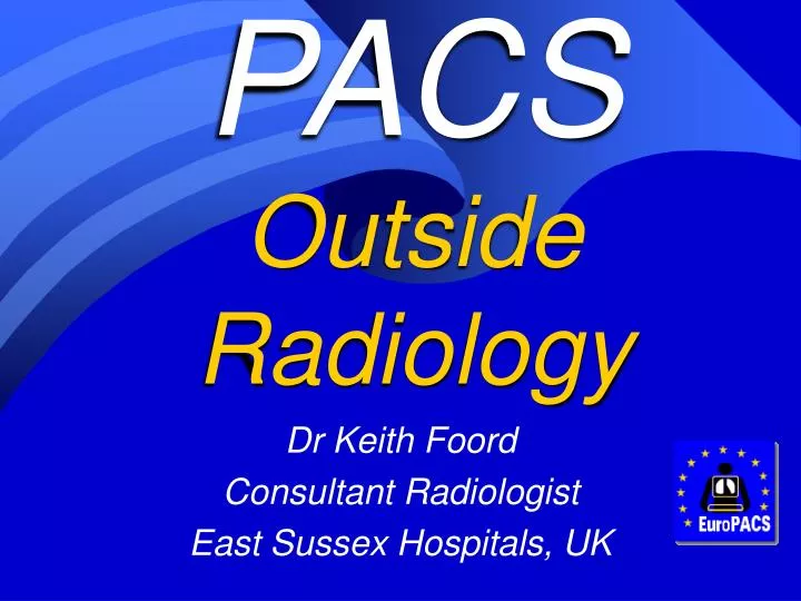
PACS Outside Radiology. Dr Keith Foord Consultant Radiologist East Sussex Hospitals, UK. `basic’ web viewers used in many clinical settings. Radiology integration in the multi-disciplinary workplace. Enterprise wide Multi-speciality PACS.

E N D
PACSOutside Radiology Dr Keith Foord Consultant Radiologist East Sussex Hospitals, UK
Radiology integration in the multi-disciplinary workplace
Enterprise wideMulti-specialityPACS “In an Enterprise PACS Radiology images account for 30% or less of image volume…. It quickly became clear that many images sources outside of radiology were not being captured, and if they were they were not available for enterprise distribution, nor linked to the electronic patient record Another problem related to workflow. There was no RIS entry – no accession number and no entry on the DICOM MWL….. Perhaps the biggest problem with expanding PACS is the lack of informatics standards in other departments. Some ophthalmology images are so proprietary that there is (essentially) no out put.” Gary Wendt U Wisconsin SCAR 2005
EnterprisePACS “One of the things we believe is that a radiology PACS, by itself, will not provide an adequate return on investment. The (enterprise) PACS must become part of the Electronic Patient Record to take advantage of electronic ordering, scheduling and reporting. To accommodate this we add 1.5 to 2 TB of storage EACH MONTH” Ramin Khorasani MD, Director of Information Management for Radiology, Brigham and Women’s Hospital, Boston
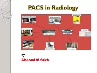
Q. Who works with images? Image producers Image viewers – passive/learning Image interpreters and reporters Image interpreters / concurrent operators Image users – planning Image users – virtual endoscopy Image users – real time guided therapy and surgery A. Almost everyone, and if they don’t access to images would enrich their clinical lives
Image producers Radiographers Nuclear Med Technicians Laboratory Technicians Medical Photographers and many others Nurse Endoscopists The IT systems they use and how they record and manage images affects the remainder of the operation……
Scanned Images Visual Light and Radiotherapy
Capturing non radiological, scanned in images (includes images of request forms if no Order-Entry system linked via an EPR) Scanned images – TWAIN High end Scanned images – ISIS Both can be converted using DICOM Secondary capture and DICOM GWL via a PACSport computer. Pdf files can now be DICOM encapsulated
Capturing Visual Light Images What is DICOM-VL? DICOM-Visible Light is an Supplement to integrate visible light images into DICOM Microscopy, endoscopy and digital photography. In particular, DICOM-VL specifies how to store preparation procedures and image-acquisition conditions in the image headers. It allows other ‘ologies’ to implement the DICOM standard, allowing their integration into EPRs It is the first real visible light image standard and allows display of these images next to radiological images on a DICOM viewing station. Part of the DICOM standard since 1999. DICOM minutes show struggle to obtain interest or expertise from medical VL companies, but better recently and DICOM MPEG2 Video soon (?now)
Pathology PACS…a rather special case! “Glass slides must be scanned at at least 400x Generates up to 6 GB per slide Scanning a single slide can take up to 20 mins, but developments are expected to reduce this to 1 minute. Compression of 15:1 can be applied, but will still generate 134 GB per day – 3 TB per month This will require 350 TB storage over 10 years” Kai Saeger, Charite Institute of Pathology, Berlin CARS2003
“In the last few years, dynamic telepathology has been the only real digital application that has been able to transmit wide areas of microscopic preparations Dynamic telepathology is a real-time technology system. Only a few systems are able to store and create a database of the transmitted cases e.g. Bliss by BacusLabs. http://www.bacuslabs.com//. A WebSlide can be created either in uncompressed .bmp format or in .jpg format. These systems are very expensive because they work by means of a robotic microscope, and the software for image viewing and interaction is often sophisticated and only available with the purchase of apparatus. A WebSlide can be created either in uncompressed .bmp format or in .jpg format” Each of these slides from one pathological specimen must be Scanned (BacusLabs system) Francesco Alfredo Zito1, Franco Marzullo1, Diego D'Errico1, Cesare Salvatore1, Rosanna Digirolamo1, Angela Labriola1 and Antonio Pellecchia2 1 Division of Pathology2 Division of Digestive Endoscopy, National Cancer Institute, Bari, Italy
This is a 3 ´ 2 mm image of an endoscopic gastric biopsy. The 7200 ´ 5200pixel image was digitized at an objective magnification of ´ 20. A total of 20 still pictures are required to generate a large image of85 MB in TIFF format, which is then compressed into a 9 MB Quick Time VR file. The entire digitization process takes 60 min. The quality of the movie is related to the quality of initial images Quicktime virtual reality technology in light microscopy to support medical education in pathology Francesco Alfredo Zito1, Franco Marzullo1, Diego D'Errico1, Cesare Salvatore1, Rosanna Digirolamo1, Angela Labriola1 and Antonio Pellecchia2 1Division of Pathology2Division of Digestive Endoscopy, National Cancer Institute, Bari, Italy
Image viewers – passive/learners As most viewers are not specialists need skilled interpretive reports with images. Means must have coherent IT systems to input these with no error. Related images – and their reports should be linked together Agfa Web 1000 Bachuslabs.com SPECT-CT PET-CT
Cardiology Potential for systems fragmentation if need to maximise integration not recognised by different project groups. A corporate knowledge deficit / lateral thinking issue
Image interpreters and reporters Radiology Microbiology Haematology Histopathology Cardiology Virology Endoscopy Cytology Nuclear Medicine Opthalmology
• A & B scan Ultrasound• Corneal Topography• Automated field analysers• Optical Coherence Tomography (OCT) Opthalmology
Reminder…. “Perhaps the biggest problem with expanding PACS is the lack of informatics standards in other departments. Some ophthalmology images are so proprietary that there is (essentially) no out put.” Gary Wendt U Wisconsin SCAR 2005
Image interpreters and concurrent operators. Use old images to plan, but dynamic image users in operation, recording new images - still and short video clips to be added to patient file Endoscopic intervention Interventional Radiology Orthopaedic Surgery Acute Interventional Cardiology
Image users – planning Oncology Orthopaedics Neuroscience Vascular Surgery Ear Nose and Throat
3D CT images – help with Ortho reconstruction CTA – diagnostic study prior to IR angioplasty of ext. iliac artery
DICOM RT is the the extension of the DICOM 3.0 standard which handles the Radiotherapy modality. It was developed to extend the current DICOM 3.0 standard to include the RT modality, rather than produce a completely new standard Seven new objects define the RT modality DICOM-RT is not a separate standard - it is an extension, defining 7 new objects:- RT image- RT dose- RT structure set- RT plan- RT treatment record (beam session, brachy session, summary). Beams are defined with a system of "control points RT ImageIncludes all the normal RT images, DRRs, portal images, simulator images and radiogramsRT DoseThe total dose distributions from the planning system; dose matrix, dose points, isodose curves and dose volume histograms.RT Structure SetPatient related structures identified from diagnostic data, CT, virtual simulations and treatment planning systems. RT PlanThis is the geometric and dosimetric data for course of external beam treatment or brachy-therapy. RT Treatment RecordRecords all the treatment session data. Radiotherapy Planning
Image users – virtual endoscopy “Virtual endoscopy focuses on the virtual representation of minimally invasive procedures for training, planning, and diagnosis without an actual invasive intervention. In the past few years, virtual endoscopy modes have been transferred from research systems in virtually every commercial medical imaging software, but with a varying quality and exibility. It is based on a 3D scan of the respective body region. Examples for these scans are CT (Computed Tomography) scans, MRI (Magnet Resonance Imaging) scans of the abdominal area, the heart, the head, the lungs, or rotational angiography of blood vessels in various body parts. Based on the resulting volumetric data, the organs of interest are visualized and inspected from interior (.endo.) viewpoints.” Virtual Endoscopy in Research and Clinical Practice Dirk Bartz, Visual Computing for Medicine Group, University of Tübingen Eurographics 2003 The video images need embedding in PACS…this video is 36 kBytes
More VIRTUAL ENDOSCOPY Planning endoscopic neurosurgery The lung tumour is not visible on virtual bronchoscopy, but the site for a trans-bronchial biopsy is mapped in green These VE images should be available in the VL endoscopy suite via enterprise PACS Virtual Endoscopy in Research and Clinical Practice Dirk Bartz, Visual Computing for Medicine Group, University of Tübingen Eurographics 2003
Image users – real time guided therapy and surgery Sinus surgery. The cross lines show The position of the probe on the corresponding CT in x,y and z axes GE Medical Systems
Digital OR Images Smith and Nephew website :UPMC Southside Hospital BrainLAB AG
HEAD-MOUNTED DISPLAYS AND AUGMENTED REALITY Visual interfaces—like haptic interfaces—are used to immerse participants in a virtual environment. These displays range from conventional desktop screens to head-mounted displays, depending on the degree of reality required. Head-mounted displays consist of goggles that afford a stereoscopic view of the computer-generated environment. A sense of motion is created by continuously updating the visual input with positional information derived from the participant's head movements. Collected by a tracking system connected to the display, this information is fed back to the computer controlling the graphics. Microvision's display system uses anatomical, biochemical, and physiological images and other data to guide surgeons through procedures. The computer assisted approach allowed a smaller, exactly placed craniotomy primarily in MCA aneurysms. 3D presentation of the aneurysms and the adjacent arteries in correct orientation facilitated identification and dissection the aneurysms. Current navigation systems are not precise enough to allow “blind” aneurysm clipping by placing a real clip on the virtual aneurysm neck. Minim Invasive Neurosurg 2003; 46: 269-277 R. Schmid-Elsaesser1, A. Muacevic1, M. Holtmannspötter2, E. Uhl1, H.-J. Steiger1 Neuronavigation Based on CT Angiography for Surgery of Intracranial Aneurysms: Primary Experience with Unruptured Aneurysms Vectorvision’s FDA Approval to market 2004 Most recent papers suggest navigational accuracy of these systems to within 1 – 1.5 mm They are not yet accuarte enough to perform say aneurysm clipping without skilled surgical final manouevres
Incorporated into the report are captured images of key findings (which can be exploded to full screen presentation), structured diagnosis information, recorded video and audio, the ability to sort findings by anatomy or priority, to view prior findings associated with the corresponding patient and hyperlinks to related information. DICOM Structured Reporting", Dr. David A. Clunie, ISBN 0-9701369-0-0 Structured reporting DICOM SR – is an ‘envelope’, but within this useful structure is available. It allows linkage to other DICOM objects, including between associated radiological and other images and waveforms. CAD outputs can be in SR format. User decides how much structure to use and controls with templates the type of content, if it is mandatory or optional and modes of expression PointDx
Dilemma – products from many different vendors – hardware & software incompatibility Goal – prevent being vendor dependent - consumer locked into specific products – component interoperability “plug and play” – seamless integration with “other” IT John A. Carrino, M.D. Assistant Professor of Radiology Information Management Group Jefferson Health System
ISSUES for Enterprise PACS • Image data set size – MDCT/Pathology • Image storage volumes • Networking • Integration • RIS becomes IIS within EPR, but preferably an integrated component of the EPR • Image and data Protocols • Lack of protocol standards and expertise with some vendors • DICOM & IHE • Corporate awareness and lateral thinking
Quite clearly if we do not all eat and think together Enterprise wide PACS may be a very long time coming
“Perhaps the biggest problem with expanding PACS is the lack of informatics standards in other departments.”Gary Wendt U Wisconsin SCAR 2005 Vendors of all medical imaging equipment, not just in radiology, must be brought into the IHE and DICOM fraternities. We need to educate hospital operators and image users in all clinical specialities not to buy bespoke IT solutions.Keith Foord Conquest Hospital UK CARS 2005 CfH looking to integrate other ‘ologies’ within 2 to 4 years