Download
1 / 46
500 likes | 662 Views
COCCIDIA (SPOROZOA ) Coccidia are members of the class sporozoa ,. The life cycle is characterized by an alternation of generations , sexual (gametogony) and asexual (schizogony) reproduction and most members of the group also share alternative hosts.

E N D
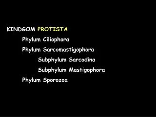
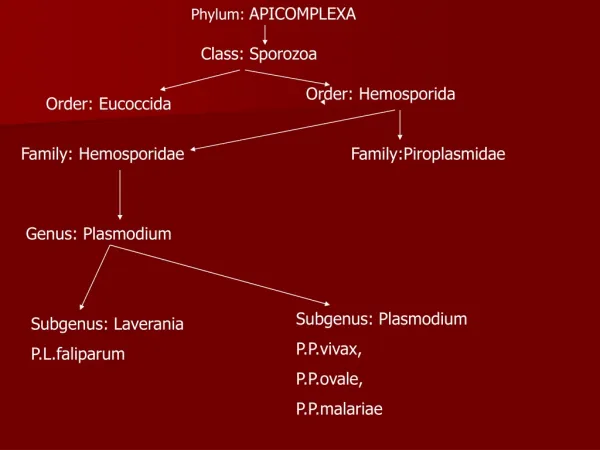
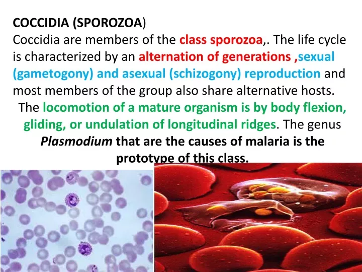
COCCIDIA (SPOROZOA) Coccidia are members of the class sporozoa,. The life cycle is characterized by an alternation of generations ,sexual (gametogony) and asexual (schizogony) reproduction and most members of the group also share alternative hosts. The locomotion of a mature organism is by body flexion, gliding, or undulation of longitudinal ridges. The genus Plasmodium that are the causes of malaria is the prototype of this class.
Plasmodium spp. disease name: Malaria There are four species normally infecting humans, namely, Plasmodiumfalciparum, Plasmodium vivax, Plasmodium ovale, and Plasmodium malariae.
Life cycle The life cycle of malaria is passed in two hosts (alternation of hosts) and has sexual and asexual stage (alternation of generations). Vertebrate host - man (intermediate host), where the asexual cycle takes place.The parasite multiplies by schizogony and there is formation of male and female gametocytes (gametogony). Invertebrate host - mosquito (definitive host) where the sexual cycle takes place.Union of male and female gametes ends in the formation of sporozoites (sporogony). The life cycle passes in four stages:
The life cycle passes in four stages: Three in man: - Pre - erythrocytic schizogony - Erythrocytic schizogony - Exo- erythrocytic schizogony One in mosquito - Sporogony
Introduction into humans - when an infective female Anopheles mosquito bites man, it inoculates saliva containing sporozoites (infective stage). 1-Pre- Erythrocytic schizogony - sporozoites reach the blood stream and within 30 minutes enter the parenchymal cells of the liver, initiating a cycle of schizogony.Multiplication occurs in tissue schizonts, to form thousands of tiny merozoites. Merozoites are then liberated on rupture of schizonts about 7th – 9th day of the bites and enter into the blood stream. These merozoites either invade the RBC’s or other parenchymal liver cells.
Note:In case of P. falciparumand possibly P. malariae, all merozoites invade RBC’s without re-invading liver cells. However, for P. vivax and P. ovale, some merozoites invade RBC’s and some re-invade liver cells initiating further Exo-erythrocytic schizogony, which is responsible for relapses. Some of the merozoites remain dormant (hypnozoites) becoming active later on.
Erythrocytic schizogony (blood phase)is completed in 48 hrs in P. vivax, P.ovale, and P. falciparum, and 72 hrs in P. malariae. The merozoites reinvade fresh RBC’s repeating the schizogonic cycles Erythrocytic merozoites do not reinvade the liver cells. So malaria transmitted by blood transfusion reproduces only erythrocytic cycle Gametogony Some merozoites that invade RBC’s develop into sexual stages (male and female gametocytes). These undergo no further development until taken by the mosquito.
Sporogony (extrinsic cycle in mosquito) When a female Anopheles mosquito vector bites an infected person, it sucks blood containing the different stages of malaria parasite. All stages other than gametocytes are digested in the stomach. The microgametocyte undergoes ex-flagellation. The nucleus divides by reduction division into 6-8 pieces, which migrate to the periphery. At the same, time 6-8 thin filaments of cytoplasm are thrust out, in each passes a piece of chromatin. These filaments, the microgametes, are actively motile and separate from the gametocyte.
The macrogametocyte by reduction division becomes a macrogamete. Fertilization occurs by entry of a micro gamete into the macro gamete forming a zygote. The zygote changes into a worm like form, the ookinete, which penetrates the wall of the stomach to develop into a spherical oocyst between the epithelium and basement membrane. The oocystes increase in size. Thousands of sporozoites develop inside the oocysts. Oocysts rupture and sporozoites are liberated in the body cavity and migrate everywhere particularly to the salivary glands. Now the mosquito is infective The sporogonous cycle in the mosquito takes 8-12 days depending on temperature
Oocysts rupture and sporozoites are liberated in the body cavity and migrate everywhere particularly to the salivary glands. Now the mosquito is infective The sporogonous cycle in the mosquito takes 8-12 days depending on temperature
Plasmodium falciparum demonstrates no selectivity in host erythrocytes, i.e. it invades young and old RBCs cells. The infected red blood cells also do not enlarge and become distorted. • Multiple sporozoites can infect a single erythrocyte, and show multiple infections of cells with small ring forms. • The trophozoite is often seen in the host cells at the very edge or periphery of cell membrane. • Occasionally, reddish granules known as Maurer’s dots are observed • Mature (large) trophozoite stages and schizonts are rarely seen in blood films, because their forms are sequestered in deep capillaries, liver and spleen. • Peripheral blood smears characteristically contain only young ring forms and occasionally crescent shaped gametocytes. Multiple sporozoites can infect a single erythrocyte crescent shaped gametocytes schizonts
Clinical features Of all the four Plasmodia, P. falciparum has the shortest incubation period, which ranges from 7 to 10 days. After the early flu-like symptoms, P.falciparumrapidly produces daily (quotidian) chills and fever as well as severe nausea, vomiting and diarrhea. The periodicity of the attacks then becomes tertian (36 to 48 hours), and fulminating disease develops. Involvement of the brain (cerebral malaria) is most often seen in P.falciparuminfection. Capillary plugging from an adhesion of infected red blood cells with each other and endothelial linings of capillaries causes hypoxic injury to the brain that can result in coma and death. Kidney damage is also associated with P.falciparummalaria, resulting in an illness called “black water” fever. Intravascular hemolysis with rapid destruction of red blood cells produces a marked hemoglobinuria and can result in acute renal failure, tubular necrosis, nephrotic syndrome, and death. Liver involvement is characterized by abdominal pain, vomiting of bile, hepatosplenomegally, severe diarrhea, and rapid dehydration.
Plasmodium vivax is selective in that it invades only young immature erythrocytes. Infections of P. vivax have the following characteristics: • Infected red blood cells are usually enlarged and contain numerous pink granules or . • The trophozoite is ring-shaped but amoeboid in appearance. • More mature trophozoites and erythrocytic schizonts containing up to 24 merozoites are present. • The gametocytes are round Plasmodium vivax P.vivax is selective in that it invades only young immature erythrocytes. Infections of P. vivax have the following characteristics: • Infected red blood cells are usually enlarged and contain numerous pink granules or schuffner’s dots. • The trophozoite is ring-shaped but amoeboid in appearance. • More mature trophozoites and erythrocytic schizonts containing up to 24 merozoites are present. • The gametocytes are round
Clinical features After an incubation period (usually 10 to 17 days), the patient experiences vague flu-like symptoms, such as headache, muscle pains, photophobia, anorexia, nausea and vomiting. As the infection progresses, increased numbers of rupturing erythrocytes liberate merozoites as well as toxic cellular debris and hemoglobin in to circulation. In combination, these substances produce the typical pattern chills, fever and malarial rigors. These paroxysms usually reappear periodically (generally every 48 hours) as the cycle of infection, replication, and cell lyses progresses. Clinical features After an incubation period (usually 10 to 17 days), the patient experiences vague flu-like symptoms, such as headache, muscle pains, photophobia, anorexia, nausea and vomiting. As the infection progresses, increased numbers of rupturing erythrocytes liberate merozoites as well as toxic cellular debris and hemoglobin in to circulation. In combination, these substances produce the typical pattern chills, fever and malarial rigors. These paroxysms usually reappear periodically (generally every 48 hours) as the cycle of infection, replication, and cell lyses progresses.
Plasmodium ovale P. ovale is similar to P. vivax in many respects, including its selectivity for young, pliable erythrocytes. As a consequence the classical characteristics include: • The host cell becomes enlarged and distorted, usually in an oval form. • Schiffner’s dots appear as pale pink granules. • The infected cell border is commonly fimbriated or ragged • Mature schizonts contain about 10 merozoites.
Clinical features The incubation period for P.ovale is 16-18 days but can be longer. Clinically, ovale malaria resembles vivax malaria with attacks recurring every 48-50 hours. There are however, fewer relapses انتكاساتwith P.ovale. Less than 2% of RBCs usually become infected.
Plasmodium malariae In contrast with P.vivax and P.ovale, P.malariae can infect only mature erythrocytes with relatively rigid cell membranes. As a result, the parasite’s growth must conform to the size and shape of red blood cell. This requirement produces no red cell enlargement or distortionتشويه, but it results in distinctive shapes of the parasite seen in the host cell, “band and bar forms” as well as very compact dark staining forms. The schizont of P.malariae is usually composed of eight merozoites appearing in a rosette.
Clinical features The incubation period for P. malariae is the longest of the plasmodia, usually 18 to 40 days, but possibly several months to years. The early symptoms are flu-like with fever patterns of 72 hours (quartan or malarial) in periodicity.
Laboratory diagnosis Microscopic examination of thick and thin films of blood is the method of choice for confirming the clinical diagnosis of malaria and identifying the specific species responsible for disease. Malaria parasites in thick and thin blood films are best stained at pH 7.1 – 7.2 using a Romanowsky stain The thick film is a concentration method that may be used to detect the presence of organisms. The thin film is most useful for establishing species identification. Serologic procedures are available but they are used primarily for epidemiological surveys or for screening blood donors.
Prevention • Chemoprophylaxis and prompt diagnosis and treatment. • Control of mosquito breeding • Protection of insect bite by screening, netting and protective clothing • Use of insect repellents.
Toxoplasma gondii – causes toxoplasmosis. The definitive host is the domestic cat and other felines. Humans and other mammals are intermediate hosts. T.gondii is usually acquired by ingestion and transplacental transmission from an infected mother to the fetus can occur. Human–to–human transmission, other than transplacental transmission, does not occur. Toxoplasma gondii – causes toxoplasmosis. The definitive host is the domestic cat and other felines. Humans and other mammals are intermediate hosts.T.gondii is usually acquired by ingestion and transplacental transmission from an infected mother to the fetus can occur. Human–to–human transmission, other than transplacental transmission, does not occur.
After infection of the intestinal epithelium, the organisms spread to other organs, especially the brain, lungs, liver, and eyes. Most primary infections in immunocompetent adults are asymptomatic. Congenital infection can result in abortion, stillbirth, or neonatal disease with encephalitis, chorioretinitis and hepatosplenomegaly.
For the diagnosis of acute and congenital infections, an immunofluorescence assay for detection of antibody is used. Microscopic examination of Giemsa–stained preparations shows crescent–shaped trophozoite. Cysts may be seen in the tissue.
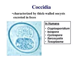
Cryptosporidium parvum– causes cryptosporidiosis, the main symptom of which is diarrhea. It is most severe in immunocompromized patients, e.g., those with AIDS. The organism is acquired by faecal-oral transmission of Oocysts from either human or animal sources. The oocysts excyst in the small intestine, where the trophozoite (and other forms) attach to the gut wall. Invasion does not occur.
The jejunum is the site most heavily infested. The pathogenesis of the diarrhea is unknown; no toxin has been identified. The disease in immunocompromized patients presents primarily as a watery, non-bloody diarrhea causing large fluid loss. Symptoms persist for long periods in immunocompromized patients, whereas self-limited in immunocompetent individuals. Although immunocompromized patients usually do not die of cryptosporidiosis, the fluid loss and malnutrition are severely debilitating.
Diagnosis is made by finding oocysts in fecal smears when using a modified acid–fast stain. Serological tests are not available. There is no effective drug therapy.