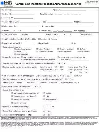
Download

1 / 4
40 likes | 373 Views
Obtaining Femoral Venous Access Annotations (E). Operator: gown, glove and mask. Risks Common--cannulation of femoral artery, line infection. Rare--injury to femoral nerve, entry into peritoneal space, retroperitoneal hematoma. Other issues: difficult to pass SG catheter without fluoroscopic gui

E N D
1. Central Line Placement Obtaining Femoral Venous Access Annotations (E)
2. Obtaining Femoral Venous Access Annotations (E) Operator: gown, glove and mask.
Risks
Common--cannulation of femoral artery, line infection.
Rare--injury to femoral nerve, entry into peritoneal space, retroperitoneal hematoma.
Other issues: difficult to pass SG catheter without fluoroscopic guidance from this position; avoid in patients with known deep venous thromboses.
Benefits
Can be placed without interrupting chest compressions in cardiac arrest
Hemorrhage can be controlled with compression.
Cannulation of artery does not require immediate removal of catheter.
3. Obtaining Femoral Venous Access Annotations (E) Positioning/Prepping
Patient should not be in Trendelenburg; ideally, should be flat and supine.
Position patient's leg in slight "frog-leg" position to open up inguinal fossa.
Prep with iodine superiorly to 10 cm above inguinal ligament, medially to scrotum or labia majora, inferiorly to 15 cm below inguinal ligament, laterally to anterior superior iliac spine.
Landmarks and Angle of Insertion: Trace the inguinal ligament from the pubic tubercule to the anterior superior iliac spine. The femoral artery lies at the junction of the medial and middle thirds of this line. The femoral pulse can be palpated just inferior to the ligament. The femoral vein lies 1 to 2 cm medial to this. The needle should be inserted 2 to 3 cm below the inguinal ligament to minimize the risk of entering the peritoneal space. (See Figure 1)
4. Obtaining Femoral Venous Access Annotations (E) Depth/Angle of Insertion: The needle should be inserted at a 45 to 60 degree angle (not a shallow angle) directed in the sagittal plane. A common error is to direct the needle in a line perpendicular to the inguinal ligament. This will cause the needle to pass medial to the vein. The vein is usually at a depth of greater than 2 cm; in obese patients the needle may need to be hubbed in order to obtain access. (See Figure 2) The fingers of the opposite hand can be positioned to help guide your needle and avoid puncturing the artery. Place the second and third fingers over the medial aspect of the femoral artery. The needle should always point in front of these fingers which are positioned over the artery in order to avoid or reduce the risk of sticking the artery.
5. Obtaining Femoral Venous Access Annotations (E) Common Problems/Fixes
Unable to palpate femoral pulse in a code situation. Accept cannulation of either artery or vein. If artery is cannulated, infuse fluids and or pressors as needed until another access is gained or circulation is restored.� Remove sheath, holding pressure, when patient more stable.
Unable to locate vein. Try repositioning leg; try ultrasound; move closer to inguinal ligament.
Strong resistance to passage of needle. Likely within the inguinal ligament. Remove needle and repeat attempt more caudally.
Vein entered but unable to pass wire. May be in superficial femoral vein or leg may be positioned poorly. Reattempt from a slightly different angle; reposition leg.
First attempt yields flash but poor blood flow. Subsequent attempts yield small amounts of blood but no flow upon aspiration. Likely a hematoma has been formed and is now being entered with subsequent sticks. Try another site or use ultrasound.