Download
1 / 38
420 likes | 721 Views
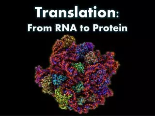
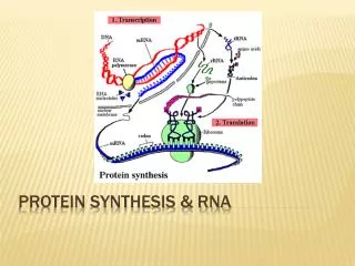
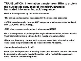
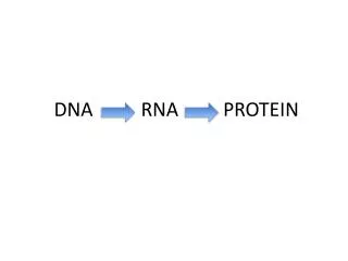
MB 207 – Molecular Cell Biology. From RNA to Protein. Translation Protein. Overall view of Protein Synthesis. The ribosome (ribosomes read messenger RNA and direct the synthesis of the encoded protein). The cellular factory responsible for synthesizing proteins

E N D
MB 207 – Molecular Cell Biology From RNA to Protein Translation Protein
The ribosome(ribosomes read messenger RNA and direct the synthesis of the encoded protein) • The cellular factory responsible for synthesizing proteins • Consists of various rRNAs and about 50 different proteins (ribosomal proteins): 2/3 RNAs & 1/3 proteins • RNAs instead of proteins with the catalytic activity, proteins are there as to stabilize the RNA core • Assembled in the nucleolus • Inactive state: exists as two subunits: a large subunit & a small subunit • When the small subunit encounters an mRNA, the process of translation of the mRNA to protein begins. • Contain 4 binding sites: 1 mRNA binding site & 3 tRNA binding sites: • A (aminoacyl) • P (Peptidyl) • E (Exit) site
Comparison procaryotic and eucaryotic ribosome structures (both have similar structure and function) S= rate of sedimentation in an ultracentrifuge
Ribosome subunits Svedberg (S): a unit of sedimentation velocity (sedimentation is an ultracentrifuge depends on both the mass and shape of a molecule
Charging the tRNA • tRNA is the translator between mRNA and protein (transfer genetic information to aa sequence) • tRNA has a specific anticodon and acceptor site • tRNA has a specific charger protein - aminoacyl tRNA synthetases • can only bind to that particular tRNA and attach the correct amino acid to the acceptor site • Energy to make this bond comes from ATP • The genetic code is translated by means of the two adaptors that act one after another: • aminoacyl-tRNA synthetase (couples an aa to it’s corresponding tRNA) • tRNA molecule (anticodons forms bp with codon on the mRNA) anticodon codon
Translation • The process that uses the base sequence in mRNA to synthesize a polypeptide with "complementary" amino acid sequence • The information in mRNA is always read from the 5' to the 3' direction. The polypeptide is synthesized from its amino terminus to its carboxyl terminus. RNA: 5'----------------------------------3' polypeptide: H2N-----------------------------COOH • The translation process occurs at the ribosome in the cytoplasm • Involves the three major classes of RNA: mRNA, tRNA, and rRNA as well as free amino acids, free energy, and several non-ribosomal protein factors
The ribosomal subunit cycle during protein synthesis Initiation factors 30S subunits with initiation factors Pool of free ribosomes Separate subunits (Ribosomal subunits assemble and disassemble during each round of protein synthesis in the ribosome cycle) Elongation Termination Initiation Initiation requires free ribosome subunits. When ribosomes are released at termination, they dissociate to generate free subunits. Initiation factors are present only on dissociated 30S subunits. When subunits reassociate to give a functional ribosome at initiation, they release the factors.
Translation initiation requires: • Ribosome brought to mRNA • Ribosome properly aligned over start codon • P site of ribosome containing charged tRNA • The initiation process differssignificantly in prokaryotesand eukaryotes • Initiation is aided by translation initiation factors
Translation initiation Bacterial mRNA are often polycistronic – encode severaldifferent proteins Eucaryotes mRNA only encode 1 single protein
Translation initiation in Prokaryotes • mRNA start codon and fMet-tRNA anticodon are aligned to start translation • Base-pairing of mRNA start codon and fMet-tRNA initiates reaction cascade to form 70S initiation complex: • IF3 dissociates • Large subuntis binds • GTP is hydrolyzed by IF2 • IF2 and IF1 dissociate • Protein synthesis begins
Initiation of Translation • The start signal for translation is the codon ATG (AUG), which codes for methionine (met) (formyl-met for prokaryotes) – all newly synthesized protein has a met at the N-terminal • A tRNA charged with met is required to bind to the translation start signal • An complex consists of the small ribosomal unit, an initiation factor (eIFs) and the tRNAmet is formed • The complex bind to the 5’ end of an mRNA (recognize the 5‘ Cap for eukaryotes or Shine-Dalgarnosequence for prokaryotes) and move forward along the mRNA to search for the initiator codon AUG - Scanning requires energy - ATP hydrolysis - It is not always the first AUG that is recognized as a start codon, sequences around first AUG might reduce efficiency of initiation so scanning continues to next AUG. • Initiation factors dissociate with GTP hydrolysis, and the large ribosomal subunit associates with the complex, the initiator tRNA bind at P site • Protein synthesis is ready to begin
The initiation phase of protein synthesis in eukaryotes • 43S pre-initiation complex formed by: • 40S subunit • Initiation factors eIFA, eIF3, eIF5B-GTP and eIF-Met-tRNA-GTP • eFI4F complex (E,G and A) binds to mRNA • 43S pre-initiation complex brought to mRNA by eIF4 family of translation initiation factors • Helicase activity of eIF4 family helps 40S subunit seat on the start codon • eIF2 and eIF3 are released from the complex • 60S subunit binds 40S subunit and initiation factors are released • Ribosome is now ready to begin protein synthesis
Elongation requires: • Placement of charged tRNA in the A site • Peptide bond formation • Translocation • Elongation factors EF-Tu, EF-G and EF-Ts
Elongation of the New Protein • A next tRNA carrying an other amino acid is attracted and pairs with the next codon at the A site, the peptide bond is catalysed by a ribosomal protein (peptidyl-transferase) associated with the large ribosomal subunit. • The result is a transfer of the N-methionine to the second tRNA in the A site which carries now a dipeptide and the initiator tRNA being uncharged in the P site. • Formation of each peptide bond is energetically favorable because the growing C-terminus has been activated by the covalent attachment of a tRNA molecule The incorporation of an amino acid into a protein
Elongation of the New Protein • The first tRNA is now released (move to E site) and the ribosome shifts so that a tRNA carrying two amino acids is now in the P site, and leaves the A site unoccupied but with the third codon exposed. • Translocation: • Ribosome moves to the next codon • Empty tRNA is ejected and the peptidyl-tRNA is shifted from the A site to the P site • New aminoacyl-tRNA is allowed to enter within the A site • Translocation is catalyzed by the elongation factor EF-G. The G indicates that this factor uses the energy gained from the hydrolysis of GTP for translocation to occur. • Finally athird tRNA is escorted by EF-Tu to the A site and the anticodon base pairs specifically with the codon. • This binding triggers the release of the initiator tRNA from the E site and allows the complex to be ready for another round of peptide bond formation, translocation and codon-anticodon base pairing • And so on…..
View of the translation cycle • The aminoacyl-tRNA is escorted to the A site by the elongation factor EF-Tu • The peptidyl transferase reaction transfer the amino acid from the P site onto the aminoacyl-tRNA in the A site Incorrectly base paired tRNAs preferentially dissociate • During translocation: • Ribosome moves one codon down mRNA • tRNA with growing protein chain moves into P site • Spent tRNA moves into E site • Translocation requires EF-G
Translating an mRNA molecule Step 1 An aminoacyl-tRNA bind to a vacant A-site on the ribosome. Step 2 A new peptide bond is form Step 3 The mRNA moves a distance of 3 nts through the small –subunit chain, ejecting the spent tRNA molecule and ‘resetting’ the ribosome so that the next incoming aminoacyl-tRNA molecule can bind The 3 steps cycle is repeated over and over again during protein synthesis.
Termination • Stop codon signals termination: UAG, UGA, UAA • Release factors (RF) accomplish termination • Termination is similar in prokaryotes and eukaryotes
Termination of the Protein Synthesis • This release in turn causes the complex to dissociate and mRNA, tRNA and ribosomal subunits are freed. • When the ribosome reaches a stop codon, no aminoacyl tRNA binds to the empty A site. This is the ribosomes signal to break into its large and small subunits, releasing the new protein and the mRNA. • Stop codons are triplets which are not recognized by any tRNA (UAA, UAG, UGA), but by a protein releasing factor (RF1 or RF2 in prokaryotes, eRF in eukaryotes). • The factor R binds to the A site forcing the peptidyl transferase to catalyze the addition of water to the peptidyl-tRNA and causes the release of the polypeptide chain The final phase of protein synthesis
Polyribosome • A single mRNA molecule is not only translated once • As soon as the ribosome has moved away from the initiation site, another round of initiation can begin • A single mRNA is often transcribed by many ribosomes at the same time, usually 100 to 200 bases apart from each other • A group of ribosomes on the same mRNA is called a polyribosome or polysome • Many proteins can be made in a given time
Protein folding • Polypeptide chain acquires its secondary and tertiary structure as it emerges from a ribosome. The N-terminal domain folds first, while the C-terminal domain is still being synthesized. • The protein has not yet achieved its final conformation by the time it is released from the ribosome. • Mechanisms that monitor protein quality after protein synthesis: 1) Correctly fold and assemble protein will be left alone 2) Incompletely folded proteins were refolded, with the help from molecularchaperones: e.g. hsp70, hsp60-like proteins 3) Incompletely folded proteins that can not be refold will be digested by proteosomes 4) Combination of all of these processes is needed to prevent massive protein aggregation in a cell
Action of chaperones during translation Chaperones bind to the amino (N) terminus of the growing polypeptide chain, stabilizing it in an unfolded configuration until synthesis of the polypeptide is completed.
Protein folding • The co-translational folding of a protein: • A growing polypeptide chain is shown acquiring its secondary & tertiary structure as it emerges from a ribosome. The hsp70 family of molecular chaperones: Recognize a small stretch of hydrophobic amino acids on a protein’s surface. Hsp70 (together with hsp40) binds to it’s target protein and hydrolyzes a molecule of ATP to ADP, undergoing a conforma- tional change result in hsp70 clamping tightly to target. Hsp 70 then dissociate induced by rapid re-binding of ATP. Repeated cycles of hsp protein binding & release help the target protein to refold. hsp70 machinery hsp70 machinery
Protein aggregate Newly synthesized protein Hsp70, 60 & 40 Correctly folded without help Correctly folded with help from chaperone Incompletely folded forms – digested in proteosome Increasing time The cellular mechanisms that monitor protein quality after protein synthesis: • Combination of processes needed to prevent massive aggregation in a cell, which can occur when many hydrophobic regions on protein clump together • and precipitate the entire mass.
Different levels of protein structure • Proteins come in a wide variety of shapes, and generally 50 to 2000 amino acids long • Primary structure: Sequence of amino acids along the core of the polypeptide chain. • Secondary structure: Folding of the polypeptide into most energetically favorable conformation resulting from various non-covalent bonds that form between one part of the chain and another. • Tertiary structure: The full 3-D organization of a polypeptide chain. • Quaternary structure: The highest level of organization, recognized in protein, formed as a complex of more than one polypeptide
Secondary Structure • Four different noncovalent bonds: ionic bond, H-bond, Van der Waals attraction & hydrophobic/hydrophilic attraction Folded conformation in aqueous environment Unfolded polypeptide
Secondary Structure • Two most common types: -helix & -sheet • -helices are held together by H-bond between the N-H and C=O groups of the polypeptide backbone 4 aa away • Form a regular helix with a complete turn of 3.6 aa
Secondary Structure • -sheets are held together by H-bond between the N-H in the peptide bond on one strand and the C=O of a peptide bond on another sheet strand • Produce a very rigid sheet structure • parallel vs anti-parallel -sheet anti-parallel parallel
Tertiary Structure • Besides the 4 non-covalent bonds, one of the important features is the formation of disulfide bonds S-S bonds (between cysteines) cysteine Interchain disulfide bond oxidants reductants Intrachain disulfide bond
Tertiary Structure • Protein domain: a substructure produced by any part of a polypeptide chain that fold independently into a compact, stable structure with a specific function, i.e. catalytic domain SH3 domain Small kinase domain SH2 domain Large kinase domain
Quaternary structure • Overall 3D structure assumed by the multimeric protein • Aggregates of more than one polypeptide chain • Individual polypeptide chains that make up multimeric proteins are often called protein subunits 4o structure of Hemoglobin
Types of protein based on structure: Globular & Fibrous Globular: • Tightly folded polypeptide chains, having a much more compact structure. • Globular proteins include most enzymes and most of the proteins involved in gene expression and regulation, e.g. hemoglobin, deoxyribonucleases, cytochrome c DNAse Cytochrome c
Types of protein based on structure • Fibrous: • Elongated structures, with the polypeptide chains arranged in long strands (parallel strands along a single axis). • Major structural components of the cell or tissue • Examples: collagen (tendon, cartilage, and bone), elastin (skin), tubulin and actin (cell shape, motility, muscle movement) Collagen triplehelix
Assembly of proteins • Proteins molecules often serve as a subunits for the assembly of large structure • Benefits: Free subunits Assembled structures • A large structure built from repeating subunits requires smaller amount of genetic information • Both assembly and disassembly can be readily controlled, reversible processes • Errors in the synthesis of the structure can be more easily avoided, since correction mechanisms can operate during the course of assembly to exclude malformed subunits dimer helix ring
Proteins • Many proteins have non-peptide components such as carbohydrate moieties (glycoproteins), metal groups (metalloproteins), lipids (lipoproteins). • Function of proteins • structure: hair, fingernails. • transport: hemoglobin. • information: protein hormones. • catalysis: enzymes. • locomotion: muscles. • Protein family: a group of proteins with members having similar - amino acids sequence - three-dimensional (3-D) structure & - function (eg. serine proteases)