Download

1 / 39
400 likes | 426 Views
MRI of the Wrist: Algorithmic Approach for Evaluating Wrist Pain. Greg S. Matthews, MD Bahram Kiani , MD Scott D. Wuertzer , MD Jason A. Powell, MD Brandon L. Roller, MD, PhD Leon Lenchik , MD Maha Torabi , MD. Authors’ Affiliation:

E N D
MRI of the Wrist: Algorithmic Approach for Evaluating Wrist Pain Greg S. Matthews, MDBahram Kiani, MDScott D. Wuertzer, MDJason A. Powell, MDBrandon L. Roller, MD, PhDLeon Lenchik, MDMahaTorabi, MD
Authors’ Affiliation: Department of Radiology, Division of Musculoskeletal Imaging Wake Forest School of Medicine Wake Forest Baptist Medical Center One Medical Center Blvd Winston-Salem, NC 27156 Corresponding Author: MahaTorabi, MD, (e-mail: mtorabi@wakehealth.edu) All authors have disclosed no relevant relationships.
Review an algorithmic approach for evaluating wrist pain. • Examine conditions of the wrist and review their characteristic MRI findings. • Correlate MRI findings with those seen at arthroscopy. • Describe the commonly used surgical classification systems that guide treatment.
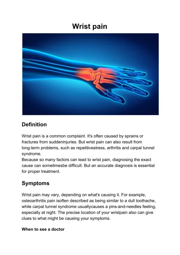
Introduction • Subacute or chronic wrist pain usually develops gradually, with or without an acute traumatic event. • The differential diagnosis is extensive and includes intra-articular and extra-articular conditions. • MRI helps narrow the differential diagnosis and guide surgical planning. a b Coronal MR image (a) and arthroscopic midcarpal image (b) of the wrist depict the lunate (L), triquetrum (T), and the scaphoid (S) bones.
Algorithmic Approach to Evaluating Wrist Pain • Using an algorithmic approach for evaluating wrist MR images, the radiologist can help the surgeon plan for treatment. • Surgical classification systems are used to guide treatment options. • Intra-articular wrist conditions may be treated with arthroscopic or open surgery. • Extra-articular wrist conditions are usually treated with open surgery.
Algorithm Used for Evaluating Wrist Pain
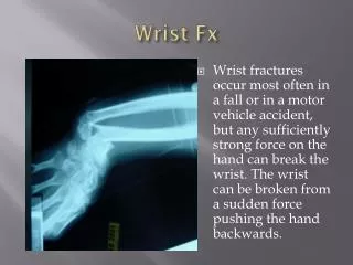
Intra-Articular: Osseous Conditions • Common intra-articular osseous conditions include: • Fractures • Avascular necrosis (AVN) • Ulnocarpal impaction • Stylocarpal impaction • These may be treated with arthroscopic or open surgery.
Case 1: Hook of Hamate Fracture Crush injury and persistent ulnar-sided wrist pain in a 23-year-old woman. Radiographs obtained 4 weeks prior to MRI were negative for fracture. MR images show subacute fracture through the hook of hamate. c b (a) Radiograph of the wrist does not show findings of an acute fracture. (b) Axial T1-weighted MR image obtained at the level of the distal carpal row shows a fracture (arrow) through the hook of hamate. (c) Axial T2-weighted fat-saturated MR image shows a fracture (arrow) with hamate marrow edema.
Kienböck Disease Management • Described by Robert Kienböck in 1910 • Multifactorial pathophysiologic mechanism • Causes: increase in shear forces on the lunate owing to anatomic factors such as negative ulnar variance, rectangular or square geometry of the lunate, vascular pattern without sufficient collateral vessels, or venous congestion • Acute trauma or repetitive minor trauma may also predispose a patient to Kienböck disease. • Historically, for assessment and staging, various classification systems (eg, Lichtman classification system) have been used. However, many surgeons are starting to use the arthroscopic classification system in symptomatic patients, as proposed by Bain et al.1
Kienböck Disease Classification • Grading is based on the number of articular surfaces affected when visualized at arthroscopy1 • Grade 0: None • Grade 1: Proximal lunate only • Grade 2a: Proximal lunate + distal radius • Grade 2b: Proximal and distal lunate with coronal lunate fracture • Grade 3: Proximal and distal lunate + distal radius • Grade 4: Grade 3 + proximal capitate Illustration depicts the various grades in the Kienböck disease classification system. Red area = surface affected. Figure reprinted and adapted, with permission, from reference 2. 0 1 2a 2b 3 4
Kienböck Disease • The proposed arthroscopic grading system can guide management. • If conservative management fails, the following procedures can be performed: • Grade 0 Radial or capitate shortening osteotomy with or without revascularization procedure • Grade 1 Proximal row carpectomy or limited carpal fusion such as radioscapholunate fusion • Grade 2A Radioscapholunate fusion • Grade 2B Proximal row carpectomy • Grade 3 or 4 Wrist fusion or wrist arthroplasty
Case 2: Grade 2B Kienböck Disease Chronic wrist pain in a 37-year-old woman. MR images show AVN, complicated by a coronally oriented fracture of the lunate. The patient underwent treatment with a proximal row carpectomy. a c b Sagittal T2-weighted fat-saturated (a) and axial T1-weighted (b) MR images obtained at the level of the proximal carpal row show AVN of the lunate with a coronal fracture (arrow). (c) AP radiograph of the wrist shows proximal row carpectomy.
Case 3: Grade 4 Kienböck Disease Gradually increasing wrist pain over 6 months, which was unresponsive to conservative management, in a 32-year-old woman. MR images show AVN with fragmentation of the lunate. (c) Arthroscopic midcarpal image shows high-grade capitate chondrosis (arrow). (d) Arthroscopic radiocarpal image shows lunate fragmentation. c c Lunate Capitate a b e Sagittal T2-weighted fat-saturated (a) and coronal gradient echo (GRE) (b) MR images show lunate fragmentation (arrow) and high-grade lunate and capitate chondrosis. (e) AP wrist radiograph shows wrist arthrodesis, with dorsal plate fixation. d d
Intra-Articular: Soft-Tissue Conditions • Common intra-articular soft-tissue conditions include: • Triangular fibrocartilage complex (TFCC) lesions: • - Traumatic Palmer 1 classification scheme • - Degenerative Palmer 2 classification scheme • Scapholunate (SL) and lunotriquetral (LT) ligament tears • Chondral lesions • Synovitis • These conditions are usually treated arthroscopically.
Palmer Classification and Treatment of Traumatic TFCC Lesions TFCC lesions without associated distal radioulnar joint instability are initially treated conservatively. If immobilization and steroid injections fail, arthroscopic intervention can be considered. The traumatic lesions are classified as: - 1A: Central disk tear (often with an unstable flap) Treated with débridementof the torn portion - 1B: Tear of ulnar insertion (+/- ulnar styloid fracture) TFCC repair - 1C: Tear extends to ulnolunate or ulnotriquetral ligaments Mini-open approach, +/- flexor carpi ulnaris augmentation - 1D: Tear of radial insertion Treated with débridementof the torn portion 1C Ulnar styloid Volar Illustration shows the various classifications of TFCC lesions according to the Palmer classification system. Figure reprinted and adapted, with permission, from reference 2. Distal radius 1B Dorsal 1A 1D +/- = with or without.
Case 4: Palmer 1B Lesion Persistent ulnar-sided wrist pain after a fall in a 58-year-old woman. MR and arthroscopic images show a TFCC tear at the ulnar styloid attachment, which was repaired. b c Arthroscopic image (c) shows a probe in the TFCC tear (arrowhead) and suture repair of the tear (arrow). Coronal GRE (a) and coronal T2-weighted fat-saturated (b) MR images show a Palmer 1B TFCC tear (arrow).
Case 5: Palmer 1D Lesion Chronic ulnar-sided wrist pain in a 57-year-old man with a history of trauma. MR images show a radial attachment TFCC tear. The tear was treated with arthroscopic débridement. a b (a, b) Coronal T1-weighted fat-saturated MR arthrograms show a tear (arrow) of the radial attachment of the TFCC.
Palmer Classification of Degenerative TFCC Lesions • Grading system: • 2A: TFCC fraying • 2B: TFCC fraying with chondrosis of the lunate or ulnar head • 2C: TFCC perforation with chondrosis of the lunate or ulnar head • 2D: Grade 2C with tear of LT ligament • 2E: Grade 2D with ulnocarpal arthritis • If conservative management fails, Palmer type 2A lesions can be treated • with open ulnar diaphyseal shortening. The Palmer 2B, 2C, 2D, and 2E lesions can be treated with TFCC débridementor repair, with the option of performing ulnar shortening osteotomy. At this time, there is controversy with no consensus on the best treatment option.
Palmer Classification of Degenerative TFCC Lesions Schematic Representation Reprinted, with permission, from reference 3..
Case 6: TFCC Tear Intermittent ulnar wrist pain for 1 year exacerbated by activity in a 54-year-old woman. MR image shows a central TFCC tear without lunate or ulnar chondrosis. Arthroscopy confirmed a degenerative TFCC tear, which was treated with débridement. * a b (a) Coronal GRE MR arthrogram shows a central TFCC tear (arrow), with extension of the radiocarpal contrast material into the distal radioulnar joint (DRUJ). (b) Arthroscopic image shows a degenerative central TFCC tear (*), which was débrided.
Case 7: Palmer 2A Lesion Chronic right wrist pain localized on the ulnar side in a 57-year-old man. MR arthrogram shows central TFCC fraying without associated chondrosis, which was débridedarthroscopically. * a b (a) Coronal T1-weighted fat-saturated MR arthrogram shows central TFCC fraying (arrow). (b) Arthroscopic image shows a shaver débridingan area of central TFCC fraying (*).
Case 8: Palmer 2D Lesion • Worsening wrist pain in a 17-year-old boy after a fall in the driveway 4 months prior. MR and arthroscopic images show a central TFCC tear and a tear of the LT ligament. a c b e d T L * Arthroscopic images show a central TFCC tear (* in c), débridementof the volar LT ligament with a shaver (d), and the LT ligament (arrow in e) following débridement. L = lunate, T = triquetrum. (a,b) Coronal T2-weighted fat-saturated MR images show a tear of the LT ligament (arrow ina) and central perforation of the TFCC (arrow in b).
Geissler Arthroscopic Classification of SL and LT Ligament Injuries • Grade 1: Ligament fraying with normal carpal alignment • Grade 2: Ligament tear with slight carpal bone step-off; cannot pass 1-mm probe through the tear • Grade 3: Ligament tear with carpal bone step-off, seen in radiocarpal and midcarpal spaces; able to pass 1-mm probe through the tear • Grade 4: Ligament tear; able to pass 3-mm arthroscope through the tear (drive-through lesion); gross instability with manipulation
Case 9: Grade 2 SL Tear Wrist pain in a 52-year-old woman. MR images show a tear in the mid-portion of the SL ligament. The probe could not be passed through the defect at arthroscopy. a b c d (b, c) Radiocarpal arthroscopy images show a tear of the SL ligament (arrow), which could not be probed. (d) Midcarpal arthroscopic image shows slight carpal step-off at the SL interval (arrow). (a) Coronal T2-weighted fat-saturated MR arthrogram shows linear perforation of SL ligament (arrow).
Case 10: Grade 3 SL Tear Two-year history of wrist stiffness, weakness, and pain in a 51-year-old woman. MR images show a volar-sided SL ligament tear with mild widening of the SL interval. a b c d Coronal T1-weighted (a) and T1-weighted fat-saturated (b) MR arthrograms show a contrast material–filled linear defect (arrow) through the SL ligament. (c, d) Radiocarpal arthroscopic images show a tear and hemorrhage in the SL ligament, which was débrided(d). e f (e, f) Midcarpal arthroscopic images show an SL interval gap (arrow in e), through which a 1-mm probe could be passed (f).
Intra-Articular: Soft-Tissue Conditions • Synovial conditions include: • Septic arthritis • Inflammatory arthropathies • Synovial osteochondromatosis • Pigmented villonodularsynovitis (PVNS)
Case 11: Septic Arthritis Chronic wrist pain related to chronic septic arthritis and osteomyelitis in a 22-year-old man with a long history of intravenous drug use. b a c d Coronal T1-weighted (a), coronal T2-weighted fat-saturated (b), axial T1-weighted (c), and axial T2-weighted fat-saturated (d) MR images obtained at the level of the carpal bones show extensive radiocarpal and midcarpal synovitis, carpal and metacarpal marrow edema, and destructive changes of the scaphoid. The findings are consistent with chronic septic arthritis and osteomyelitis. There is secondary SL advanced collapse (SLAC), with proximal migration of the capitate (arrows in a and b).
Case 12: Synovial Osteochondromatosis Gradually increasing left wrist pain and a palpable dorsal mass in a 53-year-old man. MR images show multiple osteochondral bodies, a finding consistent with synovial osteochondromatosis of the DRUJ, which was confirmed at surgery. a b c (a) AP wrist radiograph shows numerous joint bodies (arrows) in the DRUJ. Coronal GRE (b) and axial T2-weighted fat-saturated (c) MR images show effusion and synovitis in the DRUJ, with low signal intensity foci, a finding representing osteochondral bodies (arrow).
Case 13: PVNS Wrist mass along the volar aspect of the ulna in a 42-year-old woman. MR images show an intra-articular mass with susceptibility artifact on GRE images, a finding suggesting hemosiderin deposition. The findings are most consistent with PVNS, which was confirmed at surgery. c a b d (a, b) Axial T1-weighted MR image (a) and T1-weighted fat-saturated MR image obtained after the administration of contrast material (postcontrast) (b), obtained at the level of the proximal carpal row, show an enhancing soft-tissue mass (arrow) in the pisotriquetral recess. (c, d) Coronal T2-weighted fat-saturated (c) and GRE (d) MR images show the mass with low signal intensity (arrow in c) on the T2-weighted image and blooming susceptibility artifact (arrow in d) on the GRE image.
Extra-Articular: Soft-Tissue Conditions • Extra-articular soft-tissue conditions include: • Tendon injuries • Aneurysms and vascular malformations • Neuromas and nerve sheath tumors • Accessory muscles
Case 14: Traumatic Tendon Tear Complicated by Infection Increasing wrist pain and swelling 1 week after a cat bite to the dorsal wrist of a 59-year old woman. MR images show cellulitis and an abscess in the dorsal wrist and a complete tear of the extensor pollicis longus (EPL) tendon. c d a b (a) Axial T1-weighted fat-saturated postcontrast MR image obtained at the level of the distal radius shows dorsal wrist cellulitis with a small rim-enhancing collection, a finding consistent with an abscess (arrow). (b, c) Axial T2-weighted fat-saturated (b) and T1-weighted (c) MR images obtained at the level of the distal radius show an empty third extensor compartment tendon sheath (arrow) owing to a complete EPL tear. (d) Sagittal T2-weighted fat-saturated MR image shows the proximal end of the retracted EPL (arrow).
Case 15: De Quervain Tenosynovitis Pain along the radial aspect of the wrist for 6 weeks in a 21-year-old swimmer. MR images show tenosynovitis of the first extensor compartment, which was treated with a US-guided corticosteroid injection. a a b c d (c) Longitudinal power Doppler US image of the distal wrist shows tenosynovitis with synovial thickening and hyperemia of the first extensor compartment (arrow). (d) Transverse gray-scale US image shows the needle with the tip in the first extensor compartment tendon sheath for steroid injection (arrow). Sagittal T1-weighted fat-saturated postcontrast (a) and axial T2-weighted fat-saturated (b) MR images obtained at the level of the distal radius show extensive tenosynovitis (arrow) of the first extensor compartment, deep relative to the pain marker. Radius Radius
Case 16: Ulnar Bursitis Carpal tunnel syndrome in a 53-year-old woman. MR images show rim-enhancing fluid surrounding the flexor tendons, proximal and distal to the carpal tunnel, a finding representing ulnar bursitis. The symptoms resolved with conservative management. c a b Axial T2-weighted fat-saturated (a) and axial T1-weighted postcontrast (b) MR images obtained at the level of the distal radius and coronal T2-weighted fat-saturated (c) MR image show ulnar bursitis associated with the flexor tendons (arrow). The coronal image (c) also shows extension of bursitis in the palm.
Case 17: Vascular Malformation Right third finger pain and an associated mass that slowly increased in size in a 31-year-old woman. MR images show a vascular malformation in the radial aspect of the third finger, which was confirmed at angiography and subsequent surgery. (a) Coronal T2-weighted fat-saturated MR image shows a lobulated T2-hyperintense soft-tissue mass (arrow). (b) Coronal T1-weighted maximum intensity projection postcontrast-subtracted MR image shows an arterial phase microlobular enhancement pattern (arrows). (c) Frontal digital subtraction angiographic image obtained after brachial artery injection shows a vascular malformation (arrows). a b c
Case 18: Traumatic Neuroma Right wrist pain, weakness, and numbness in a 42-year-old man with history of trauma. MR images show an enhancing mass along the course of the median nerve. a b c FPL FDS FCR Axial T1-weighted (a), T1-weighted fat-saturated (b), and T1-weighted fat-saturated postcontrast (c) MR images obtained at the level of the distal forearm show an enlarged enhancing median nerve, consistent with a neuroma (solid arrows) between the flexor digitorum superficialis (FDS), flexor carpi radialis (FCR), and flexor pollicis longus (FPL) tendons (dashed arrows in a). A normal caliber median nerve was identified proximally, and perineural scarring was found distal to the neuroma.
Case 19: Accessory Muscle Palpable mass in the hypothenar eminence in a 37-year-old mechanic, which was painful when the patient did push-ups. MR images show an accessory abductor digitiminimi muscle overlying the ulnar nerve, which may be associated with ulnar nerve compression. b a (a, b) Axial T1-weighted MR images show an accessory abductor digitiminimi muscle (arrow), deep relative to the marker indicating the site where the symptoms manifest.
Conclusion • An algorithmic approach, combined with a knowledge of common wrist conditions, will help the radiologist assess the wrist on MR images. • With a familiarity of the surgical classification systems and the expected management of various wrist conditions, the radiologist can describe clinically relevant findings depicted on MR images. • The radiologist can then convey this pertinent information to the referring clinicians, thereby improving patient care.
Arnaiz J, Piedra T, Cerezal L, et al. Imaging of Kienböck disease. AJR Am J Roentgenol 2014;203(1):131–139. • Bain GI, Begg M. Arthroscopic assessment and classification of Kienbock’s disease. Tech Hand Up Extrem Surg 2006;10(1):8–13. • Chloros GD, Wiesler ER, Poehling GG. Current concepts in wrist arthroscopy. Arthroscopy 2008;24(3):343–354. • Rominger MB, Bernreuter WK, Kenney PJ, Lee DH. MR imaging of anatomy and tears of wrist ligaments. RadioGraphics 1993;13(6):1233–1246; discussion 1247–1248. • Torabi M, Martell B, Tuohy C, Lenchik L. MRI-arthroscopy correlation of the wrist: a primer for radiologists. Current Radiology Reports. 2016;4(2):1-3. • Zanetti M, Saupe N, Nagy L. Role of MR imaging in chronic wrist pain. Eur Radiol 2007;17(4):927–938.
References • Bain GI, Begg M. Arthroscopic assessment and classification of Kienbock’s disease. Tech Hand Up Extrem Surg 2006;10(1):8–13. • Torabi M, Martell B, Tuohy C, Lenchik L. MRI-arthroscopy correlation of the wrist: a primer for radiologists. Current Radiology Reports. 2016;4(2):1-3. • Cockenpot E, Lefebvre G, Demondion X, Chantelot C, Cotten A. Imaging of sports-related hand and wrist injuries: sports imaging series. Radiology. 2016;279(3):674-92.