Download

1 / 47
470 likes | 701 Views
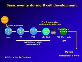
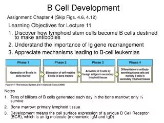
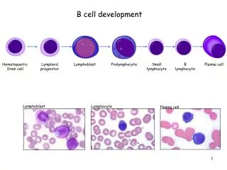
Micro 297 Graduate Immunology Lecture 15 Development of B Lymphocytes I Generation of Antibody Diversity Thursday August 14, 2003 Michael Wolcott READING Chapter 7. Overview of B-Cell Development. Generation of Lymphocyte Antigen Receptor.

E N D
Micro 297 Graduate ImmunologyLecture 15Development of B Lymphocytes IGeneration of Antibody DiversityThursday August 14, 2003Michael WolcottREADING Chapter 7
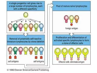
Generation of Lymphocyte Antigen Receptor • The expression of the antigen receptor is the defining (and essential) event in the development of both B and T cells. • Antigen receptors, in the form of Ig’s on B cells and the TCR on T-cells, are the means by which lymphocytes sense the presence of antigen in their environment. • The diverse repertoire of lymphocyte receptors is accomplished through complex and elegant genetic mechanisms. • The basic mechanism for generation of diversity is common to both B cells and T cells and involves many if not all of the same enzymes.
Recall these Facts • The receptors produced by each lymphocyte have a unique antigen specificity which is determined by the structure of their antigen-binding site. • The wide range of antigen specificities in the antigen receptor repertoire is due to variation in the amino acid sequence in the V region. • Each individual possesses billions of lymphocytes, these cells collectively provide the individual with the ability to respond to a great variety of antigens. • In each chain the V region is linked to an invariant constant region which provides effector function
Genetic Model Compatible with Ig Structure Any model must accommodate known properties of Ig’s • The vast diversity of antibody specificities • The presence of a variable region and a constant region • The existence of different isotypes with the same antigen specificity • Germline Theory • Each antibody specificity coded by a germline (inherited) gene • What are the problems with this theory? • Somatic Mutation Model • Genome contains a small number of genes from which the diversity of antibody specificities is generated by mutation. • What are the problems with this theory?
Problems with the Germline and Somatic-Mutation Models • Germline Theory • Part of gene subject to wide variation and part characterized by relative constancy. • So many specificities – so few genes • Same variable regions on different isotypes • Somatic Mutation Theory • Part of gene subject to wide variation and part characterized by relative constancy. • Same variable regions on different isotypes • Resolution Begins • Dreyer-Bennent Hypothesis
The Dreyer – Bennett Hypothesis Two genes – one polypeptide chain • Two separate genes encode a single Ig H or L chain • One gene for the V region and a separate gene for the C region • The two genes come together at the DNA level and are transcribed together • Thousands of V genes and a single C gene • Strength of this Recombination Model • Accommodates one part of the molecule varying with the other part remaining relatively constant • Accommodates how a single V region can be associated with more than one isotype • PROBLEM • So many specificities - so few genes!
Southern Blot Proof of Dryer-Bennett Hypothesis Ig genes are rearranged in B cells Conclusion • Embryonic cells have the genes encoding the V region and C region considerably separated in the genome. • During B cell development the genes are for V and C regions are brought much closer together. • This simple experiment showed that segment of genomic DNA within the Ig genes are rearranged in cells of the B-lymphocyte lineage, but not in other cells.
Each V Region is Encoded by More Than One Gene Segment • Cloning and sequencing of Ig genes showed even greater complexity that predicted by Dreyer and Bennett. • The DNA sequence encoding a complete V region is generated by the somatic (site specific) recombination of separate gene segments. • A single C gene segment encodes the C region
Gene Segments of VL and VH Regions • Light chain V region – two gene segments • V gene segment –first 95-101 amino acids • J (joining) gene segment – up to 13 amino acids • Heavy chain V region – three gene segments • V and J gene segments • D (diversity) gene segment
There are Multiple Different V-region Gene Segments • The immunoglobulin gene segments are organized into three cluster or genetic loci – the κ, λ, and heavy-chain loci – each on a separate chormosome. • The V gene segments can be grouped into families in which each member shares at least 80% sequence identity with other in the family. • The families can be grouped into clans, made up of familes that are more similar to each other than to families in other clans. • VH gene segments identified from amphibians, reptiles, and mammals fall into three clans. • This suggests that these clans existed in a common ancestor of these modern animal groups.
V-region Genes are Constructed From Gene Segments by Somatic (Site Specific) Recombination • VL by V-J recombination producing VJ variable region gene. • VH by D-J recombination followed by V to D-J recombination producing VDJ variable region gene.
Genetic RecombinationGeneral vs. Site Specific • Genetic recombination • the ability of DNA to undergo rearrangement that can vary the particular combinations of genes present in any individual genome. • General recombination • genetic exchange that takes place between any pair of homologous DNA sequences, usually located on two copies of the same chromosome – e.g. the exchange of sections of homologous chromosomes in the course of meiosis. • Site specific recombination • DNA homology is not required. Instead, exchange occurs at short, specific nucleotide sequences that are recognized by site-specific enzymes – examples: integration of lambda phage in bacteria and somatic recombination of gene segments in Ig’s and TCR’s.
Rearrangement of V, (D), and J gene segments is guided by flanking sequences Recombination Signal Sequences – RSS – ensure that DNA rearrangements take place at the correct location relative to the V, D, or J gene segment • Conserved heptamer and nonamer sequences flank the gene segments • The spacer between the hepatmer and nonamer sequences is always approximately 12 bp or approximately 23 bp. • The spacer varies in sequence but its conserved length corresponds to one or two turns of the DNA double helix. This brings the heptamer and nonamer sequences to the same side of the DNA helix • The heptamer – spacer – nonamer is called a recombination signal sequence - RSS • 12/23 rule – recombination, for the most part, only occurs between a 12 bp (one turn) and a 23 bp (two turn) RSS
The reaction that combines V, D, and J gene segments involves both lymphocyte-specific and ubiquitous DNA-modifying enzymes The complex of enzymes that act in concert to effect somatic V(D)J recombination is termed the V(D)J recombinase • The products of the two genes rag-1and rag-2 (recombination-activating genes) comprise the lymphoid-specific components of the recombinase. • The remaining enzymes in the recombinase are ubiquitously expressed DNA-modifying proteins that are involved in DNA repair, DNA bending, or the modification of the ends of the broken DNA. These include DNA ligase, DNA-dependent protein kinase (DNA-PK), and Ku which is a heterodimer (Ku 70:Ku 80) that associates with DNA-PK.
V(D)J Recombination is a Multistep Process • RAG protein complexes bind to 12 and 23 bp spaced RSS • The protein complexes bind to each other bringing together the segments to be joined • The DNA is cleaved to create a hairpin structure at the ends of the Ig gene segments • Other DNA-modifying proteins bind to the hairpins and the cleaved RSS ends • The DNA hairpins are cleaved at random. Additional bases may be added by TdT or subtracted by exonuclease to generate imprecise ends. • DNA ligase IV joins the ends of the gene segments to for the coding joint and the RSS ends to form the signal joint.
Intervening DNA “looped out” and lost at the next cell division Recombination of gene segments that are in the opposite orient-ation results in inver-sion and integration of the intervening DNA Light Chain Recombination Can Occur By Either “looping out” or “Inversion” Which mechanism utilized depends on the orientation of the V and J segments. Opposite Orientation “Inversion” Same Orientation “looping out”
Are RAG-1 And RAG-2 the only lymphoid specific enzymes necessary for recombination of Ig or TCR gene segments? YES So how would you prove it?
Summary of the experimental identification of RAG-1 and RAG-2 • Retroviral construct containing a promoter sequence,V and J gene segments with flanking RSS and a gene that confers resistance to mycophenolic acid (in the opposite orientation of the promoter). • The orientation of the RSS sequences requires that rearrangement occurs by inversional recombination. • If the construct is rearranged then the resistance gene is brought into the same orientation as the promoter and the cells become resistant to mycophenolic acid. • A variety of cells were tested with this system and only pre- B cells and pre-T cells were able to rearrange the V and J segments. • However, fibroblasts could carry out the rearrangement if transfected with DNA coding RAG-1 and RAG-2.
Productive vs. Non-productiveRearrangements • The joining of the V(D)J gene segments is imprecise and gene segments can be joined out of phase, thus the triplet reading frame for translation is not preserved. • In such a non-productive rearrangement, the VJ or VDJ unit will contain numerous stop codons, which interrupt translation. • When joined in phase, the reading frame is preserved and thus is a productive rearrangement.
Generation of Antibody Diversity • Multiple germline gene segments • Combinatorial V-(D)-J joining • Junctional flexibility • P-region nucleotide addition (P-addition) • N-region nucleotide addition (N-addition) • Combinatorial association of light and heavy chains. • Somatic hypermutation
Summary: The combination of many sources of diversity generates a vast repertoire of antibody specificities from a limited number of genes Diversity with in the Ig repertoire is achieved by several means. • V regions are encoded by separate gene segments, which can be brought together by somatic recombination to make a complete V region gene. • Many V region gene segments are present in the genome, thus providing a heritable source of diversity. • Combinatorial diversity results from the random recombination of separate V , D and J gene segments to form a complete V region exon. • Variability at the joints is increased by N-region and P-region additions and by the variable deletion of nucleotides at the ends of coding sequences. • The association if different light and heavy chain V regions to form the antigen-binding site of an Ig molecule contributes further to the diversity. • Finally, after an immunoglobulin is expressed, the coding regions of the V regions are modified by somatic hypermutation following stimulation of the B cell by antigen.
Structural Variation in Immunoglobulin Constant Regions • The immunoglobulin H-chain isotype are distinguished by the structure of their constant regions. • Antibody C-regions confer functional specialization. • Co-expression of IgM and IgD on B cells results from alternatively spliced H-chain transcripts. • Transmembrane and secreted forms of Ig’s are generated from alternative H-chain transcripts. • CLASS SWITCHING – the same VH exon can associate with different CH genes in the course of an immune response.
Organization Of The Ig Heavy-chain C-region Genes In Mice And Human
Co-Expression of IgD and IgM • Mature B cells that co-express IgM and IgD on their surface have not undergone class switching. – instead: • In mature B cells, transcription initiated at the VH promoter extends through both Cµ and Cδ exons. • The long primary transcript is then processed by cleavage and polyadenylation (AAA), and by splicing. • In this process there is no alteration at the DNA level. • The differential processing of the long mRNA transcripts is developmentally regulated. Immature B cells express only IgM; mature B cells IgM and IgD; activated B cells lose expression of IgD and express a single isotype of Ig. • The exact function of IgD on the surface of mature B cells is unclear. Gene-target mice lacking the delta exon appear to have normal immune responses.
Transmembrane and Secreted Forms of Ig’s Both forms are derived from the same H-chain gene sequence • Each H-chain has: • Two exons that encode the transmembrane region and the cytoplasmic tail • One exon that encodes the carboxy-terminus of the secreted form • The events that dictate whether a H-chain RNA will result in secreted or transmembrane occur during processing of the initial transcript. • The selection of transmembrane or secreted form is developmentally regulated. Prior to antigen stimualtion B cells make predominately the transmembrane form. However, plasma cell make exclusively the secreted form.
Isotype Switching Involves Recombination Between Specific Switch Signals
Isotype Switching • Repetitive DNA sequences that guide isotype switching are found upstream of each of the C-region genes. • Switching occurs by recombination between these repetitive sequences (switch signals). • Isotype switching results in deletion of the intervening DNA • Since the intervening DNA is deleted back switches are not possible, but additional switches to down stream isotypes is possible • The initial switching event takes place from the µ switch region • Subsequent switches to other isotypes take place from the recombinant switch region formed after µ switching. • Isotype switching is unlike V(D)J recombination is several ways • All isotype switching is productive • It uses different recombination signal sequences and enzymes • It happens after antigen stimulation not during B cell development • The switching process is not random – it is regulated by external signals from T cells
Isotype Switching Involves Recombination Between Specific Switch Signals
Somatic Hypermutation Somatic hypermutation further diversifies the Ab repertoire • Introduces variation into the rearranged immunoglobulin V-region that is subject to positive and negative selection • Occurs in the germinal center following antigen stimulation of the B cell. • Somatic hypermutation requires signals from activated T cells • Hypermutation is thought to occur due to the introduction of double strand breaks in the DNA of V regions, followed by error prone repair. • Hypermutation occurs at a similar time to class switching, but appear to involve different enzymes and mechanisms. • We will cover hypermutation in more detail in the lectures on B cell activation.
Regulation of Ig-Gene Transcription • Immunoglobulin genes are expressed only in B cells • Genes are expressed at different rates during different stages of development • Three major classes of cis regulatory sequences in DNA regulate transcription of Ig-genes • Promoters – short nucleotide sequences extending about 200 bp upstream from the start site, that promote intiation of RNA transcription – orientation dependent. • Ehancers – nucleotide sequences siturate some distance upstream or downstream from a gene that activate transcription from the promoter sequence in an orientation-independent manner • Silencers – nucleotide sequences that down-regulate transcription, operating in both directions over a distance