Download
1 / 44
440 likes | 551 Views
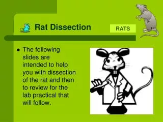
Dissection of the Rat. An Exploration of the Various Organs Systems. General Rules. No Food or Drink at your Station at all times Participation pts. Will be lost!!! Remain at your station at all times unless instructed Keep track of all instruments (do a count everyday) No open toed shoes

E N D
Dissection of the Rat An Exploration of the Various Organs Systems
General Rules • No Food or Drink at your Station at all times • Participation pts. Will be lost!!! • Remain at your station at all times unless instructed • Keep track of all instruments (do a count everyday) • No open toed shoes • Do not cut anything unless instructed • Always follow your Dissection guide
Dissection Tasks • Researcher & Photographer: • Sets up computer (obtains images & info.) • Wipes down the entire table table top • Takes photographs, prints & shares with group • Corrects all IQ’s for the group • Researches key information • Keeps the group in check with Dissection Manual • Equipment manager: • Cleans, dries, and returns all supplies to the appropriate places. • Organizes & counts all intruments • Assists with the Clean up procedures (pan, table, etc) • Makes labels/prepares them on pins • Dissectors/Assistant( min. 2 people) - Observe the dissection procedures and share with rest of the group.
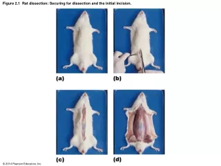
Initial Procedures to Begin the Dissection • Dissection Guide: turn to the appropriate Pgs. Beginning through pg. 16 so you can answer the first two set of Guiding Questions. • Collect all the Instruments & place them in a pouch that you have labeled with your period and group members. • Freezer Bag: using a sharpie label your bags as follows with your period and all lab group member. • Choose a rat and give it a name. Choose both a male and female name for now. You will verify the gender later and circle that name on the bag then. • Use the rat to help answer some of the guiding questions. • Tie one piece of string on the upper extremities with each end on each forelimb and another string on the lower extremities (you will keep these strings tied on the rat at all times.
Closing Procedures at the End of Class • Obtain some paper towels and roll the rat in them then place your rat into the labeled freezer bag. • Make a TIGHT Seal with all the air evacuated out. • Clean your tray and instruments. • Put all your instruments in you pouch or box. • Place tray, instruments, and rat in your designated drawer. **Spray down the ENTIRE table top with windex and thoroughly clean it leaving no streaks. **FOLD your Lab coats & stack neatly in the back Return the dissection guides and computer back to the cart. **You will store your Pan, Instruments, and Rats in the available drawers located along the sides of the room.
Instruments • 1 dissecting pan • 1 Lg. Freezer Bag • 4 pieces of string (25 in each) • 2 forceps (fine & blunt) • 1 scapel (pick up later) • 2 blunt probes • 4 sharp probes • 14 pins (6 large & 8 fine) • 2 scissors (large & fine point) • Water dropper & cup • Ruler (pick up later) • Index card (cut into labels) – need to bring some next week)
Day 1: Guiding Questions #1 • Make a list of instruments you’ll be using and the number of each. • After reading the objectives of the lab, how many organs for each system are you required to know and identify. • Name the two major regions of the integumentary system. Name three functions of this system. • Name the four key parts to the skeletal system. What does Hemopoiesis mean? • What is the only function of the muscular system? Name the three parts of the body mentioned that the muscular system plays a role. • Name the three parts of the nervous system. Which parts make up the central nervous system? • Name two functions of the endocrine system. What chemical is produced by the glands that make up this system? How is this system different from the nervous system? Name the eight types of organs that make up this system.
Guiding Questions #2 • What purpose does the circulatory system serve? What is it primarily composed of? • Name the six key structures that make up the respiratory system. What purpose does this system serve? Name the minute little sacs in the lungs that help with gas exchange. • Name the six parts of the digestive system. Where does digestion begin? Which part of the system is used to remove water from the food? • What roles do the pancreas and liver play in the digestive process? • Name one of the key waste products excreted by the Urinary system. Name the four main organs of this system. • Name the key structures of the male and female.
GuidingQuestions #3 1. From your dissection guide which chapter discusses the muscular system? • Before identifying the muscles the rat must be skinned. The first incision to be made begins on the -surface of the _____ and continues down to the point just anterior to the ______. • Circumvent cuts are made to the (right,left) by the genetials and mouth. How does the guide suggest that you separate the skin from the body? • Before separating the muscles and identifying them you first need to remove the tough white connective tissue that surrounds the muscles. Name this tissue. • Using Figure 14 (lateral view) list all of the muscles labeled in the diagram. You will ID these in your rat first.
Basic Procedure • Ventral Incision & Extension to the limbs • Separating skin from the underlying muscles • Separate the fascia off the muscle • Identify the indicated muscles -Make labels for structures listed on overhead
GuidingQuestions #3 1. From your dissection guide which chapter discusses the muscular system? • Before identifying the muscles the rat must be skinned. The first incision to be made begins on the ______ -surface of the _____ and continues down to the point just anterior to the ______. • Circumvent cuts are made to the (right,left) by the genetials and mouth. How does the guide suggest that you separate the skin from the body? • Before separating the muscles and identifying them you first need to remove the tough white connective tissue that surrounds the muscles. Name this tissue. • Using Figure 14 (lateral view) list all of the muscles labeled in the diagram. You will ID these in your rat first.
Separate & ID these Muscles (10 muscles) -Masseter -Sternomastoideus -Spinodeltoideus -Latissimus dorsi -Triceps -Brachialis -Biceps femoris -Gluteus Maximus -Tensor fascia Latae -Semitendinosus Please use the following in your guide: Lateral View (Pg. 16, Fig 14)
Muscles to locate & ID from Ventral view (14 muscles) -Pectoralis major -Biceps brachii -Pronator teres -Flexor carpi radialis -Palmaris longus -Triceps -Pectoralis minor -Rectus abdominus -External oblique -Sartorius -Gracilis -Gastrocnemius -Vastus medialis -Semimembranosus
Guiding Questions #4(Ch. 4 Digestive system) • Name the three pairs of salivary glands commonly found in the rat. • Within the mouth cavity if you were to look inside you would observe a hard and soft palate. Where are they specifically located? • After opening the abdominal cavity you should be able to locate the esophagus and follow it to the stomach. Name two structures that would be located next to the stomach. • On page 28 of your guide sheet the diaphragm separates two cavities. Name them. • How many lobes is the liver is divided into? Name them. • Name the three regions of the stomach. How is the great omentum different from the lesser omentum? • Where is the spleen located with reference to the stomach? • Name all of the structures discussed from #11 to #19.
Lg. Intestine Cecum Ascending colon Transverse colon Descending colon Sigmoid colon Oral cavity Mouth Tongue Pharynx Salivaryglands Esophagus Sm. Intestine Duodenum Jejenum Ileum (ileocecal valve) Liver Stomach Pyloricsphincter Stomach Gall-bladder Smallintestine Pancreas Smallintestine Largeintestine Rectum Figure 21.4 Anus
Bile Liver Stomach Gall-bladder Acid chyme Bile Duodenum ofsmall intestine Pancreas Figure 21.10A
Large Intestine Reclaims Water Largeintestine(colon) • Undigested material passes to the large intestine, or colon • Water is absorbed • Feces are produced Endof smallintestine Small intestine Rectum Anus Nutrientflow Appendix Cecum Figure 21.11
Guiding Questions #4(Ch. 4 Digestive system cont’d) Tasks to still complete: Finish separating the intestinal tract from the mesentery Transect the intestine from STOMACH end off the esophagus -Measure the length of the intestines (____ cm.) -Make a cross section of the stomach and make observations -Label all key structures
Guiding Questions #5(Ch. 5 Circulatory system-Pgs 31-36) • What was injected into the veins and arteries of your rat? Why was this done? • The pericardial membranes surround the . Name the two types of membranes present. How are these two membranes different from each other? • Which area (chamber) does blood enter the heart? Name the vein that drains blood from the lungs to the left atrium. • Name the structure that divides the two ventricles of the heart. • How many branches come off the aortic arch? Name them. • Where are the following arteries found? -coronary, carotid, subclavian, hepatic, gastric, femoral, caudal and intercostal? • Is the jugular a vein or artery? • What is the difference between an artery and a vein? Which one moves blood away from the heart?
Tasks to complete for the Circ. System • Find the FIVE major vessels off the aorta • Cross section of the heart • Trace major blood vessels in the arms and legs
See pgs 569-575 Pulmonaryartery 11. vessel Aorta 10. vessel Pulmonaryartery 1. vessel Superiorvena cava LEFTATRIUM 2. chamber RIGHTATRIUM Pulmonaryveins Pulmonaryveins 9. vessels 3. valve Semilunarvalve Semilunarvalve 8. valve Atrioventricularvalve 7. valve Atrioventricularvalve 4. vessel Inferiorvena cava 5. chamber RIGHTVENTRICLE 6. chamber LEFTVENTRICLE Figure 23.4A
RBC Pathway through the Circulatory System Blood from Systemic Circuit Vena cava (inferior & superior) Right atrium (Tricuspid valve-AV valve) Right ventricle (Pulmonary semilunar valve) Pulmonary circuit –Lungs (P. arteries LungsP. veins) Left atrium (Bicuspid “Mitral” valve) Left Ventricle (Aortic semilunar valve) Aorta (arch, coronary, carotid, & abdominal, renal, mesenteric, iliac arteries)
GuidingQuestions #6 • Name the five branches that come off the aortic arch. • How many chambers does the heart have? (After splitting the heart in two). Name these chambers. • Find the following blood vessels in your rat and label them: Carotid artery, Pulmonary artery, Vena Cava, Aorta, Renal, and iliac arteries (left & right). • Name the gland that sits on top of the kidney. (see Chapter IV. • The kidney are said to be “retroperitoneal” what does this mean? • Name the small tubular structure that comes out of the kidney and into the urinary bladder
Tasks to Complete for Urinary System • Trace the three main structures that come to and from the kidney • Trace the ureters from the kidney to the bladder • Locate adrenal glands • Cross section of the kidney
GuidingQuestions #7 • When looking within the kidney, the outer layer is called the __________ ________ and the inner layers is called the ________ __________. • What function does the kidney serve? • Where does the ureter transport urine to? How many ureters are there? • Name the gland that sits on top of the kidney. • Identify each structure as either male or female: -Ovary -Cowpers gland -Vas deferens -Uterus -Prostate gland -Epididymas • An oviduct is the same thing as a ________ _______
Tasks to Complete for Reproductive System • Determine sex • Find (2) major structures to verify the sex
GuidingQuestions #8 • Name the three layers of the meninges that covers the brain. • The cerebrum is composed of two hemispheres separated by a longitudinal groove called the ________ _________. • Name the nerve fibers that connect the two hemispheres that is easily viewed by a sagital section of the brain. • A highly convoluted structure found on the posterior part of the brain is called the __________. • The most primitive and most posterior part of the brain that tapers into the spinal cord is called the __________ ___________. • How many ventricles (cavities) are found in the brain? • Name the gland that sits within the sella turcica. • How many pairs of cranial nerves emerge out of the brain? Name 5 of these cranial nerves shown in Fig 27.
Be sure you have the following labels and pin these on your rat Set #1 (abdominal cavity)Set #2 (Neck & Thoracic cavity) -Liver (4 lobes) -Heart Heart (atrium, ventricle, septum) -Stomach -Aortic arch -Small intestine (D, J, I segments) -Jugular veins -Spleen -Trachea -Kidney (right) -Thyroid gland -Aorta (abdominal) -Lung (5 lobes) -Iliac arteries -Thymus gland -Diaphragm -Renal artery & vein -Adrenal gland -Carotid arteries -Mesentery -Pericardial membrane -Ureter -Inferior Vena Cava -Large intestine (caecum) -Coronary (artery or vein) **** Final Set: Ovary & uterus OR testes, vas deferens
Practice/Review Practicum • Obtain your rat and label (3) structures (A, B, & C) for the region I Assign your table. • Be sure to make write your labels clearly with pin pierced in the structure Station #1: Lateral muscles Station #2: Ventral Muscles Station #3: Neck Station #4 & #5: Thoracic Cavity Station #6 & #7: Abdominal cavity Station #8: Reproductive Structures (male) Station #9: Reproductive Structures (female) ** Number a blank sheet of paper from #1-27 (you’ll turn this sheet on Monday) **At the conclusion (10 min prior) you will need to: Clean up your table -Turn in your rats -Turn in all your instruments, placing them in the designated area in the back -Spray & clean your drawers