Download
1 / 31
310 likes | 791 Views
Ellipsoid tessellation algorithm for modelling fruit microstructure. H K Mebatsion. Why modelling fruit microstructure?. “We have to investigate the smallest details in order to appreciate the wider spectrum.” “ Multiscale Modelling” Fruit microstructures are a web of interconnected cells

E N D
Ellipsoid tessellation algorithm for modelling fruit microstructure H K Mebatsion
Why modelling fruit microstructure? “We have to investigate the smallest details in order to appreciate the wider spectrum.” “ Multiscale Modelling” • Fruit microstructures are a web of interconnected cells • Microstructures are the building block of the macrostructure • Yet, microstructures are not homogeneous • Elegant modelling procedure not affected by heterogeneity is a challenge
Microstructural heterogeneity c b a What you see is what you can see a) cortex c) v-c transition b) vascular bundle
2D microstructural modelling • Voronoi tessellation (Mebatsion et al.,2006a) Porosity=15.95% Apple parenchyma CVD PVD Porosity=15.51% Porosity=16.17%
Finite element simulation (Greenstar) Cell Pore O2 concentration profile Tri Ho (unpublished)
Deeper investigation of microstructure Middle lamella Individual cells Cell wall Cells Cells a b ESEM image of Granny Smith, Nieto et al., 2004 Synchrotron image (C. Pear) c d TEM images of Jonagold (c) and conference pear (d)
Cell Intercellular space Cell wall • Ellipse tessellation (Mebatsion et al.,2006b) Porosity= 16% Porosity=17%
Investigation of O2 profile at the microscale Conference pear Tri Ho (unpublished)
3D microstructural modelling Minimum Volume Circumscribing Ellipsoid (MVCE) • Cells in 2D are elliptical. Should they be ellipsoidal in 3D? • How to determine the geometrical properties of ellipsoids (size, position…)? • Is there something like ellipsoid fitting algorithm in 3D?
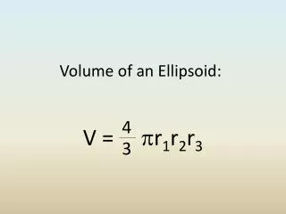
MVCE • Given points in 3D, there a unique ellipsoid with a minimum volume that encloses these sets of point • An ellipsoid can be given by the following standard equation n (1)
Then the equation of ellipsoid in the centre form reduces to (1.1)
We want points to be inside the ellipsoid hence, they must satisfy And the volume of the ellipsoid, is given by the volume of unit sphere (1.2) (2)
Hence, the optimization problem is all about • To guarantee the positive volume (avoid trivial solution) • lift the set of points • Each point is lifted to a hyperplane
The MVCE is determined as • The solution is obtained using Khachiyan's algorithm • The optimization problem is converted into concave optimization problem • Lagrangian dual approach
MVCE algorithm ( Conditional Gradient Ascent Method) N Y initialization
Previous Minimum Volume Circumscribing Ellipsoid (MVCE) algorithms took into account all sets of points • In our approach, we determine the MVCE of a polytope determined by the Convhull of the set of points • Our approach is many times faster!!!
Comparison of previous and our approaches 101 points 45 points 2.2 times as fast
Evaluation of MVCE code (2D) MVCE Centroid =[101.4004 152.66] l1=161.8438 l2= 252.3563 Ellipse fitting Centroid =[101.3835 153.8091] l1=167.8051 l2= 271.0583
Evaluation of MVCE code (3D) MVCE Centroid =[0 0 0] R1=R2=R3=1 Centroid =[0.3887, -0.2099, -0.0954] * 1.0e-016 R1=R2=R3=1
Implementation of MVCE • Image acquisition Synchrotron experiment
Implementation… • 120 phase contrast images were used • Digitization of cells in different slices is impractical • Digitization at every 10th slice (compromised) • Getting 3D coordinates of individual cells • MVCE calculation of individual cells • Visualization Visualization tool
Results MVCE MVCE
Results MVCE
Comparison of our approach and Amira ThreeDview version 1.1 Conference pear Amira version 4.2
Ellipsoid Tessellation algorithm Start MVCE (i) Non-overlap region (i) Y MVCE (j) Overlap ? N (NOR) + (MVCE (i)) MVCE (i) Y N Y Intersection of regions N End
Ellipsoid tessellation • A brand new approach Volume distribution Pear parenchyma tissue
Ellipsoid tessellation • A brand new approach Volume distribution Porosity=9.3% Pear dermal tissue
Conclusion and future plan • A very fast MVCE for the 3D visualization of cells was developed • Due to the nature of the synchrotron image automatic contour detection was not possible • A brand new ellipsoid tessellation algorithms was developed • These geometries will be exported to finite element/ volume codes (Ansys)
Nothing amuses more harmlessly than computation… Mike L Klien