Download
1 / 35
360 likes | 505 Views
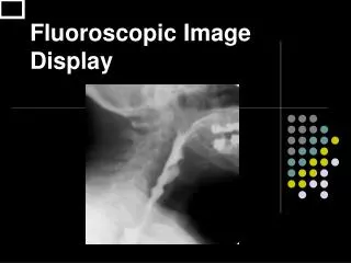
Comparison of Film v. Digital Image Display. Process of data capture. All image recording systems rely on differential absorption within the patient to produce a radiographic image Each system analog (film/screen) versus digital (CR and DDR) records photon intensity and displays the data.

E N D
Process of data capture • All image recording systems rely on differential absorption within the patient to produce a radiographic image • Each system analog (film/screen) versus digital (CR and DDR) records photon intensity and displays the data. • Differences in photon density become radiographic contrast
Radiographic Contrast • Radiographic contrast is dependent on subject contrast and technical factors • Subject contrast is basically fixed for each patient. Within certain body/organ systems we can alter subject contrast by introducing contrast media. • Some technical factors include; • kVp • Grid ratio • Screen speed • Processor temperature • Altering technical factors can improve or degrade radiographic contrast.
Film • Films produce an analog image that allows edges to gradually blend into one another. This is how we see images normally. Conversely, digital images are broken up into pixels (tiny squares). Because the image consists of pixels there is an inherent loss of spatial resolution when compared to analog film. • 2.5 – 5 lp/mm versus 10 lp/mm
Computed Radiography • Photostimulable plate (PSP) • Barium-fluorohalide doped with europium • When the PSP is struck by a photon electrons are released in the crystal lattice and stored in F centers within the lattice • In the CR reader, a helium-neon laser (red light) stimulates the crystals of the PSP. Once stimulated the crystals emit blue-violet light.
The amount of light emitted is proportional to the amount of radiation absorbed by the PSP. • The emitted light is read by a photomultiplier tube within the CR reader producing an electronic signal. The electronic signal is then fed through an analog-to-digital converter (ADC usually 12 bit) producing a digital signal. • Essentially, the PSP is a OSL radiation monitor
ADC • While converting the analog signal to a digital the ADC also breaks the pieces or squares that represent pixels in the final image.
Digital Radiography (DR) • Direct DR • X-ray photons are absorbed by the DR plate. The active ingredient is usually amorphous selenium. The a-Se is ionized by the photons and the released electrons are stored in the TFT (thin film transistor) array. The TFT consists of multitude of individual elements that represent the pixels of the final image
Indirect DR • Similar to direct DR but x-ray photons are converted to light photons (cesium iodide) and then pass through a photo-multiplier tube coupled with amorphous silicon. • Cesium iodide crystals resemble needles in appearance resulting in very little light spread and high spatial resolution.
Indirect DR Deterioration of image quality due to light diffusion
Film/screen • Optical density ranges from 0 to 3 • This represents a range of 3 orders of magnitude or 1000. • View boxes can only display 30 shades of gray • Film/screen grayscale is 1000
CR • Four orders of magnitude are possible • 10000 shades of gray • CT and MR • 12 bit or 4096 • CR and DR • 14 bit or 16384 • Digital Mammography • 16 bit or 65536 shades of gray
Contrast Resolution • Principal descriptor • Grayscale • Dynamic range
Window level • Window and leveling of the digital image allows us to see the entire grayscale of the digital image. Even though the eye is limited to 30 shades of gray by manipulating the window width and level we can determine where the 30 shades occur. • We determine the center point of the 30 shades, level, and the width is how many grays will be displayed.
Processing the Image • Image pre-processing • Find the pertinent image (histogram) • Scale data to appropriate range • Contrast enhancement • Anatomy specific gray scale manipulation • Spatial frequency enhancement
Raw Image • Inherent subject contrast displayed • Contrast inverted • PSL signal amplitude log amplified
Preprocessed raw image Scaled and inverted: Unprocessed image
Contrast Enhancement • Optimize image contrast via non-linear transformation curves • Unprocessed images have linear ‘subject contrast’ • Gradiation processing – Fuji • Tone scaling – Kodak • MUSICA - Agfa
Scaled and inverted: Unprocessed image Contrast enhanced
Spatial Resolution and Monitor Performance • In the analog environment, spatial resolution is a affected by SID, OID, RSV, FFS, and type of film to name a few. • In the digital domain, the viewing monitor also affects spatial resolution as well as contrast resolution or dynamic range.
Monitor Factors Affecting Image Quality • Monitor brightness • The brighter the monitor the better the image quality • Color v/ B & W • For CR and DR color monitors do not offer enough dynamic range • Matrix size • Most PC monitors are 1024 x 1280 or 1200 x 1600. These will allow you to view CT and MR at resolution.
Diagnostic Monitors • Matrix size • 2048 x 2560 at minimum • Black and white not color • Brightness • 600 cd/m2 v 300 cd/m2