Download
1 / 58
580 likes | 587 Views
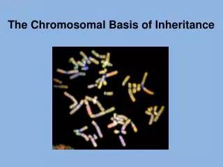
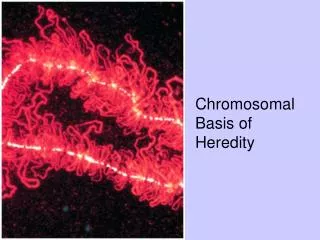
3 The Chromosomal Basis of Heredity. Chromosomes. The chromosome complement = the complete set of chromosomes of plants and animals The nucleus of each somatic cell contains a fixed number of chromosomes typical of the particular species

E N D
Chromosomes • The chromosome complement = the complete set of chromosomes of plants and animals • The nucleus of each somatic cell contains a fixed number of chromosomes typical of the particular species • The number of chromosomes vary tremendously among species and have little relationship to the complexity of the organism
Chromosomes • The chromosomes in the nuclei of somatic cells are usually present in pairs. For example, humans have 23 pairs of chromosomes • Cells with nuclei of this sort, containing two similar sets of chromosomes, are called diploid
Chromosomes • The germ cells, or gametes, are haploid and contain only one set of chromosomes, consisting of one member of each of the pairs • The haploid gametes unite in fertilization to produce the diploid state of somatic cell • The chromosomes are present in pairs because one chromosome of each pair derives from the maternal parent of the organism and the other from its paternal parent
Mitosis • Mitosis is a precise process of nuclear division that ensures that each of two daughter cells receives a diploid complement of chromosomes identical with the diploid complement of the parent cell • Mitosis is usually accompanied by cytokinesis, the process in which the cell itself divides to yield two daughter cells
Cell Cycle • In a cell that is not undergoing mitosis, the chromosomes are invisible with a light microscope. This stage of the cell cycle is called interphase • DNA in the chromosomes is replicated during a period of interphase called S = DNA synthesis • Before and after S, there are periods, called G1 and G2, respectively • These three interphase periods are followed by mitosis, M
Stages of Mitosis • Prophase is marked by the condensation of chromosomes. Each chromosome is already longitudinally double, consisting of two subunits called chromatids • Each pair of chromatids is the product of the duplication of one chromosome in the S period of interphase • The chromatids in a pair are held together at a specific region of the chromosome called the centromere.
Stages of Mitosis • At the beginning of metaphase, the mitotic spindle forms • The spindle is a bipolar structure arching between the centrosomes that consists of microtubules • The spindle fibers attach to each chromosome in the region of the centromere at the kinetochore • The chromosomes move toward the center of the cell until all the kinetochores lie on an imaginary plane equidistant from the spindle poles = themetaphase plate
Stages of Mitosis • In anaphase, the centromeres divide longitudinally, and the two sister chromatids of each chromosome move toward opposite poles of the spindle • Once the centromeres divide, each sister chromatid is regarded as a separate chromosome in its own right.
Stages of Mitosis • In telophase, a nuclear envelope forms around each compact group of chromosomes, nucleoli are formed, and the spindle disappears • The chromosomes undergo decondensation until they are no longer visible as discrete entities • The two daughter nuclei assume a typical interphase appearance • The cytoplasm of the cell divides in two
Meiosis • Meiosis is a mode of cell division in which cells are created that contain only one member of each pair of chromosomes • Meiosis consists of two successive nuclear divisions • Meiosis results in four daughter cells, each genetically different and each containing one haploid set of chromosomes • Meiosis is a more complex and considerably longer process than mitosis and usually requires days or even weeks
Meiosis • In animals, meiosis takes place in specific cells called meiocytes • The oocytes form egg cells and the spermatocytes form sperm cells • In the females of both animals and plants, only one of the four products develops into a functional cell (the other three disintegrate)
Meiosis • In plants, the products of meiosis form spores, which undergo one or more mitotic divisions to produce a haploid gametophyte organism • The gametophyteproduces gametes by mitotic division of a haploid nucleus • Fusion of haploid gametes creates a diploid zygote that develops into the sporophyte plant, which undergoes meiosis to produce spores and so restarts the cycle
Outline of Meiosis • Prior to the first nuclear division, the members of each pair of chromosomes become closely associated along their length • The chromosomes that pair with each other are said to be homologous chromosomes • Each member of a pair of homologs consists of a duplex of two sister chromatids joined at the centromere. The pairing of the homologous chromosomes therefore produces a four-stranded structure
Outline of Meiosis • At the time of pairing, the homologs can exchange genes which results in chromosomes that consist of segments from one homolog intermixed with segments from the other • In the first nuclear division, the homologous chromosomes are separated from each other, one member of each pair going to opposite poles of the spindle • Two nuclei are formed, each containing a haploid set of duplex chromosomes
Outline of Meiosis • The second nuclear division resembles a mitotic division, but there is no DNA replication. • At metaphase, the chromosomes align on the metaphase plate, and at anaphase, the chromatids are separated into opposite daughter nuclei • The net effect of the two divisions is the creation of four haploid nuclei, each containing the equivalent of a single sister chromatid from each pair of homologous chromosomes
Mitosis vs. Meiosis • Meiosis produces four cells: each contains one copy of each pair of homologous chromosomes = genetically haploid • Mitosis produces two cells which contain both members of each pair of homologous chromosomes = genetically diploid
MeiosisI • The first meiotic division—reductional division, reduces the chromosome number by half • Prophase I is the longest stage, and is commonly divided into five substages: leptotene, zygotene, pachytene, diplotene, and diakinesis These are descriptive terms that indicate the appearance of the chromosomes at each substage
Meiosis: Prophase I • Leptotene - the chromosomes first become visible as long, thread-like structures • Zygotene- synapsis of homologous chromosomes = bivalent • Pachytene - crossing-overbetween homologs • Diplotene- chromosome repulsion, however, they remain held together by cross-connections resulting from crossing-over. Each cross-connection, called a chiasma is formed by a breakage and rejoining between nonsister chromatids • Diakinesis- maximum chromosome contraction Fig. 3.9b
Meiosis: Metaphase I • Metaphase I - The bivalents positioned with the centromeres of the two homologs on opposite sides of the metaphase plate • As each bivalent moves onto the metaphase plate, its centromeres are oriented at random with respect to the poles of the spindle • Genes on different chromosomes undergo independent assortment because nonhomologous chromosomes align at random in metaphase I
Meiosis: Anaphase I • Anaphase I - homologous chromosomes, each composed of two chromatids joined at an undivided centromere, separate from one another and move to opposite poles of the spindle • The physical separation of homologous chromosomes in anaphase I is the physical basis of Mendel’s principle of segregation
Meiosis: Telophase I • Telophase I—a haploid set of chromosomes consisting of one homolog from each bivalent is located near each pole of the spindle • The spindle breaks down, the chromosomes enter the second meiotic division after only a limited uncoiling • Chromosome replication never takes place between the two divisions
Meiosis II • The second meiotic division (meiosis II) is called the equational division because the chromosome number remains the same in each cell before and after the second division • In some species, the chromosomes pass directly from telophase I to prophase II without loss of condensation • After a short prophase II and the formation of second-division spindles, the centromeres of the chromosomes in each nucleus become aligned on the central plane of the spindle at metaphase II
Meiosis II • In anaphase II, the centromeres divide and the chromatids of each chromosome move to opposite poles of the spindle • Once the centromere has split at anaphase II, each chromatid is considered a separate chromosome • Telophase II is a transition to the interphase condition of the chromosomes in the four haploid nuclei, accompanied by division of the cytoplasm.
Meiosis • The chromatids of a chromosome are usually not genetically identical because of crossing-over associated with the formation of chiasmata during prophase of the first division
Chromosome Structure • Eukaryotic chromosomes are highly coiled stable complexes of DNA and protein called chromatin • Each eukaryotic chromosome contains a single DNA molecule of enormous length • Some of the proteins present in chromatin determine chromosome structure and the changes in structure during the cell cycle • Other chromatin proteins appear to have important roles in regulating chromosome functions
Chromatin Structure • The nucleosome is the basic structural unit of chromatin • Each nucleosome is composed of a core particle, ~55 base pairs of DNA called linker DNA that links adjacent core particles and one molecule of histone H1 that binds to the core particle and to the linker DNA • Histones are small proteins that are highly conserved among different organisms
Chromatin Structure • Each core particle consists of an octamere of pairs each of histone H2A, H2B, H3, and H4; a segment of DNA containing about 145 base pairs Fig. 3.15a
Chromatin Structure • Nucleosomes coil to form higher order DNA structure called the 30-nm chromatin fiber • In the nucleus of a nondividing cell, chromatin fibers form discrete chromosome territories • Chromosome territories are correlated with gene densities • Territories of chromosome domains that are relatively gene rich tend to be located toward the interior of the nucleus
Chromatin Structure • The spaces between the chromatin domains form a network of channels large enough to allow passage of the molecular machinery for replication, transcription, and RNA processing • Replication takes place in small discrete regions that exhibit a reproducible temporal and spatial pattern, and transcription takes place in a few hundred discrete locations • The metaphase chromosome is a hierarchy of coiled coils
Chromatin Structure • Compact and heavily stained regions of chromatin are known as heterochromatin which mainly consists of highly repeated non-coding DNA sequences—satellite DNA • The rest of the chromatin, which becomes visible only after chromosome condensation in mitosis or meiosis, is called euchromatin • The number of genes located in heterochromatin is small relative to the number in euchromatin
Chromosome Structure • The centromere is essential for chromosome segregation • The centromere is a specific region of the eukaryotic chromosome. It serves as a central component of the kinetochore the complex of DNA and proteins to which the spindle fibers attach and move the chromosomes in both mitosis and meiosis
Chromosome Structure • The telomere is essential for the stability of the chromosome tips • Due to the nature of DNA replication, chromosomes require special mechanism to restore DNA in telomeres in each cycle of replication • The mechanism relies on an enzyme called telomerase
Chromosomes and Heredity • Chromosome Theory of Heredity: Genes are located in chromosomes • One of the earliest evidence that genes are located on chromosomes was found by Thomas Hunt Morgan in 1910 • Morgan’s studied inheritance patterns in Drosophila melanogaster and foundthat in some cases reciprocal crosses yield different results
Morgan’s Fruit Fly Experiments • Morgan realized that it might happen if the alleles for some genes were present in the X chromosome • The X chromosome is transmitted in a different pattern by males and females, and the Y chromosome does not contain alleles homologous to genes on the X chromosome
Nondisjunction • Experimental proof of the chromosome theory of heredity came from nondisjunction • Nondisjunction = chromosomes fail to separate (disjoin) and move to opposite poles of the division spindle, results in loss or gain of a chromosome • Calvin Bridges demonstrated that exceptional behavior of chromosomes is precisely paralleled by exceptional inheritance of their genes