Download
1 / 246
2.47k likes | 2.6k Views
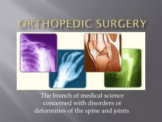
LMCC Orthopedic Review Lecture April, 2004 “Back to Basics” Dr. P.R. Thurston. Syllabus. 1. Diagnosis, Treatment & Complications of Fractures /Dislocations. 2. Diagnosis & Treatment of Arthritis. 3. Assessment and Management of Low Back Pain. &. Fractures. Dislocations.

E N D
LMCC Orthopedic Review Lecture April, 2004 “Back to Basics” Dr. P.R. Thurston
Syllabus 1. Diagnosis, Treatment & Complications of Fractures /Dislocations. 2. Diagnosis & Treatment of Arthritis. 3. Assessment and Management of Low Back Pain.
& Fractures Dislocations
Fractures Definition :- A discontinuity in the structural integrity of a bone. A fracture occurs because the force applied exceeds the breaking strength of the bone so that the Load can no longer be transferred across that zone of the bone.
Fractures All fractures ultimately begin with kinetic energy, released by misadventure and applied to the human body. Some of that energy is absorbed and some is transmitted to the surroundings. Absorbed energy must be dissipated, ie. distributed, through the soft tissues and bones. Fractures occur when the bone can not dissipate all of the energy absorbed.
Fractures Thus :- 1 ) A fracture occurs when the energy transferred to a bone exceeds the ability of the bone to dissipate that energy. 2 ) Further energy dissipation produces :- - comminution. - soft tissue damage (open fractures). - displacement. - other fractures.
Definitions Fracture:-A discontinuity in the structural integrity of a bone. Infraction:- An incomplete fracture. Dislocation:- Complete loss of contact of the articular surfaces of a joint. Subluxation:- Non-concentric joint surfaces. Reduction:- Returning a fracture or dislocation to an anatomical alignment. Comminution:-Multiple fragments.
Fractures Mechanical Properties of Bone Bone is a two-phase material :- Calcium HydroxyApatite Ca10(PO4)6(OH)2 = mineral Osteoid Collagen type I and III = fibrous Calcium is strong in compression, but weak in tension. Osteoid is strong in tension, but weak in compression.
Fractures BUT :- (for adult bone) Calcium is stronger in compression than Osteoid is in tension And therefore :- Bone always fails first in tension
Fractures For immature bone, this effect is reversed. The Buckle or Torus fracture occurs because the bone fails in compression first. In children, the Osteoid is stronger than the Mineral phase. Generally, the dislocation in youth becomes the fracture in the adult.
Fractures A bone consists of three areas :- the Diaphysis the Metaphysis the Epiphysis. Each region has its own fracture characteristics.
Fractures Oblique Diaphyseal Bending Torque Direct Traction Compression Intra-articular Pediatric Spiral Transverse Metaphyseal Epiphyseal Mixed
Bending Fractures Bending produces a transverse fracture line, with or without a lip. When load is added, the lip becomes a butterfly fragment. With more loading, the fracture line becomes oblique.
Torque Fractures - Rotatory shear produces a continually changing line of failure, giving the typical “Bayonet Spikes” at the ends of the bones. • The greater the load the longer the fracture. • These occur only in long bones and are referred to as:- ‘Spiral Fractures’
Torque Fractures $piral The butterfly segment is different from the oblique bending fracture. $
Fractures If no butterfly, then the ends are Bayonet in appearance.
Direct Blow Fractures “tapping fractures”. Fractures of “dying momentum”. Tension produced on the opposite side of the bone. Comminution produced on the impact side of the bone. High energy injuries.
Direct Blow Fractures Transverse Fractures Comminution on the opposite side to a bending fracture, ie. at the point of impact. “The Nightstick Fracture”.
Metaphyseal Fractures Traction – Avulsion. The Metaphysis is subject to all of the diaphyseal patterns plus:- 1) Traction – Avulsion. 2) Compression.
Metaphyseal Fractures Traction-Avulsion are transverse since the tension is equal on both sides of the bone. are caused by ligament or tendon traction. always occur adjacent to joints.
Fractures Traction – Avulsion.
Compression Fractures Crush fractures Impacted fractures Usually comminuted Usually axial skeleton - Vertebrae - Calcanei
Epiphyseal Fractures The Epiphysis is subject to all of the diaphyseal and metaphyseal patterns plus:- 1) Intra-articular Fractures. 2) Pediatric Fractures about the Epiphyseal plate.
Epiphyseal Fractures Intra-articular Fractures • Always require accurate reduction. • Usually require surgical treatment. • Are often comminuted. • Frequently threaten Post-traumatic Osteoarthritis.
Epiphyseal Fractures Pediatric Epiphyseal Fractures • Produce fracture patterns specific to children. • Always require accurate reduction. • Can produce growth abnormalities. • Salter-Harris Classification.
Salter-Harris Classification Fractures I II III IV V
Salter-Harris Classification Fractures 1) Fractures interfering with growing bones. 2) Worse prognosis with increasing number. 3) Probability of surgery increases with number.
Fractures A fracture can occur in :- normal bone subject to abnormal forces. = Traumatic Fractures. abnormal bone subject to normal forces. = Pathologic Fractures. normal bone subject to cyclic forces. = Fatigue or Stress Fractures.
Fractures Description 1) Displacement - Angulation 2 ) Closed or Open. 3 ) Simple or Comminuted. 4 ) Fracture Pattern eg. Spiral, Transverse etc. 5 ) Anatomical Area. 6 ) Mechanism.
Fracture Description Thisfracture is angulatedlaterally, since it points laterally. The distal fragment is tilted medially
Description Medially Displaced Closed Comminuted Short Oblique Fracture of the Proximal Humerus Caused by a direct fall
Fracture Description 1) The distal fragment is always described with relation to the proximal segment. 2) Displacement = Translation of bone ends. 3) Angulation = Orientation of bone ends. 4) Angulation identifies to where the fracture points. 5) For clarity, the tilt of the distal fragment is often used to describe angulation.
The Periosteal Bridge The Periosteal Bridge is intact on the concave side of the fracture. Reversal of the mechanism of the fracture tightens the bridge and stabilizes the fracture.
The Periosteal Bridge Tightening the periosteal bridge locks the fracture together. Holding the bridge tight requires three point fixation. “It takes a bent cast to produce a straight bone” J. Charnley
Treatment Closed or Open ( Surgical ). - Both require an understanding of fracture healing. - Closed requires reversal of mechanism of injury.
Indications for Closed Reduction There is significant displacement. Reduction is possible. The reduction, if gained, can be held. The fracture has not been produced by a traction force.
Indications for Open Reduction 1 ) There is a significant Displacement. 2 ) Open Fractures. 3 ) Intra-articular Fractures. 4 ) Un-reducible Fractures 5 ) Reductions that cannot be maintained in a cast. 6 ) Comminuted or Segmental Fractures. 7 ) Floating Joints. 8 ) Fractures with Neurovascular damage.
Open Fractures Classification :- 1. < 1 cm., inside-out, little soft tissue damage. = low potential for infection. 2. 1 cm. – 10 cms., outside-in, requires debridement, but no flap or skin graft. = moderate potential for infection. 3. > 10 cms., outside-in, high energy, devitalized muscle, comminution or bone loss, soft tissue loss.
Open Fractures Classification :- 3A. No loss of soft tissue cover, no flap required. 3B. Flap required due to soft tissue stripping. 3C. Associated vascular injury.
Degloving Mechanism Degloving Mechanism
Type III C Injuries – Vascular Injury Note pallor of the ankle No pulses
Fracture Complications 1. Pulmonary Fat Emboli 2. Compartment Syndromes 3. ‘Cast Disease’ 4. Stress Fractures 5. Pathologic Fractures
Pulmonary Fat Emboli :- A.R.D.S. - Long bone fractures, burns, contusions. - Interstitial pneumonitis due to free fatty acids - S.O.B. & confusion in young adults. - Axillary & Subconjunctival Petechiae. - Serum lipase elevated. - pAO2 reduced – if < 50 – 20% mortality. - Ventillatory support - Dexamethazone. - 5 day course.
Compartment Syndromes - increased interstitial tissue pressure. - fractures, burns, tight dressings. • normal pressure < 25 mm. Hg. • when the tissue pressure > venous capillary pressure, but less than the arteriolar pressure. • 5 P’s - pain. - pallor. - pulselessness. - paresthesias. - paralysis.
Compartment Syndrome • Symptom: Pain out of proportion to that • expected for the injury. • Signs: 1. Loss of function of muscle due to • ischemia within the compartment. • 2. Pain with passive stretch • 3. Numbness etc. are LATE findings! • 4. If neuro symptoms present, potential • for full neuro recovery is only 10 %
Rx Compartment Syndrome Release all compressive dressings / plaster. Elevate extremity to heart level. Fasciotomies.
Rx Compartment Syndrome Increased girth. Pallor of the foot. Recent surgery.