Download
1 / 86
920 likes | 1.23k Views
Skeletal system. Your bones manufacture blood cells. Our bones are held by our muscles The smallest bones are in our ears. Muscular system. Muscles are bundles of cells and fibers. We have 600 major muscles. We have 240 muscles that have specific jobs. TMJ. Structure of Bone.

E N D
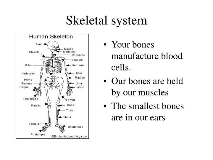
Skeletal system • Your bones manufacture blood cells. • Our bones are held by our muscles • The smallest bones are in our ears
Muscular system • Muscles are bundles of cells and fibers. • We have 600 major muscles. • We have 240 muscles that have specific jobs
Anatomy of a Long Bone • Diaphysis • Medullary Cavity • Nutrient Art & Vein • 2 Epiphyses • Epiphyseal Plates • Epiphyseal Art & Vein • Periosteum • Outer: Dense irregular CT • Inner: Osteoblasts, osteoclasts • Does not cover epiphyses • Attaches to bone matrix via collagen fibers • Endosteum • Osteoblasts, osteoclasts • Covers trabeculae, lines medullary cavity
Synovial Joint pg 215
Bursae & Tendon Sheaths • Bursae: flat, fibrous sac w/synovial membrane lining • Tendon Sheaths: elongated bursae that wraps around tendons • 3 Factors in Joint Stability: • Muscle Tone • Ligaments • Fit of Articular Surface pg 219
pg 224 Joint Shapes • Hinge: cylindrical end of 1 bone fits into trough shape of other • angular movement-1 plane (eg) elbow, ankle, interphalangal • Plane: articular surface in flat plane • Short gliding movement • (eg) intertarsal, articular processes of vertebrae
pg 225 Joint Shapes • Condyloid: egg-shape articular surface + oval concavity • side-to-side, back+forth movement • (eg) metacarpophalangeal (knuckle) • Pivot: round end fits into ring of bone + ligament • rotation on long axis • (eg) prox. radius/ulna, atlas/dens
Anterior Skull frontal bone glabella supraorbital foramen infraorbital foramen zygomatic bone mandibular symphysis maxillary bone alveolar fossa mental foramen mandible
Anterior Skull nasal bone perpendicular plate superior orbital fissure middle nasal concha inferior nasal concha bone vomer bone
Paranasal Sinuses frontal sinus ethmoid sinus maxilary sinus sphenoid sinus
frontal bone Cranium coronal suture parietal bone sagittal suture lambdoidal suture occipital bone
Ventral Skull palatine process sphenoid bone palatine bone vomer bone styloid process temporal bone mastoid process external occipital protuberance occipital bone
Occipital bone carotid canal jugular foramen occipital condyle foramen magnum
squamosal suture Lateral Skull lacrimal bone temporal bone external acoustic meatus mandibular condyle In mandibular fossa (TMJ joint)
Lateral Skull zygomatic arch sphenoid bone coronoid process sutural bone mastoid process styloid process ramus angle body mandible
cribriborm plate crista galli Internal Skull lesser wing greater wing optic canal sella turcica intenal acoustic meatus jugular foramen
Hyoid + external acoustic meatus temmporal mandibular joint Hyoid bone
________ ________ ________ Sagittal
Coronal Lambdoid Squamous
Overview of Skull Geography • Facial bones form the anterior aspect • The cranial bones enclose the brain
Vault • The cranial vault or calvaria forms the superior, lateral, and posterior aspects of skull • The cranial base forming the inferior aspect of skull
Cranial Base • Cranial base forms the skull’s inferior aspect • Three prominent ridges divide the base into fossae • The brain rests on these cranial fossae completely enclosed by the cranial vault • The brain occupies the cranial cavity
Cranium • The 8 cranial bones include; 2 parietal, 2 temporal frontal, occipital, sphenoid, ethmoid • Cranium is self- bracing allowing the bones to be thin, yet strong
Occipital bone • Forms most of the posterior wall and base of skull • Articulates with parietal & temporal • Joins w/ sphenoid in the cranial floor • Forms internal walls of posterior cranial fossa
Occipital bone - Int. landmarks • Hypoglossal canal, Posterior cranial fossa
Temporal Bone • Forms the infero-lateral aspects of the skull • Parts of the cranial floor • Divided into four regions; squamous tympanic, mastoid, and petrous-(int)
Temporal Bone • The internal petrous region contributes to the cranial base • The petrous region and the sphenoid bone form the middle cranial fossa
Temporal Bone - landmarks • Zygomatic process • Meets the zygomatic bone • Forms the cheek • Mandibular fossa • Receives condyle of mandible
Temporal bones - landmarks • Stylomastoid foramen • exit for facial nerve • Carotid canal • entrance for the carotid artery which supplies blood to cerebral hemispheres
Sphenoid bone • Bone spanning the width of middle cranial fossa • Articulates as central wedge of all cranial bones • Consists of central body and three processes; greater and lesser wings and pterygoid process (pos. view)
Sphenoid - landmarks • Sella turcica (enclosure for pituitary gland) • Optic foramina (passage of optic nerves) • Superior orbital fissure (Nerves III, IV, V enter orbit) • Foramen rotundum & ovale (Cranial Nerve V to face) • Foramen spinosum (Middle meningeal artery)
Ethmoid bone • Forms most of the area between the nasal cavity & orbits of eyes • Lies between nasal bones & sphenoid • Complex shape gives rise to nasal septum, sinuses and cribiform plate
Ethmoid bone - landmarks • Cribiform plates • Forms roof of nasal cavity • Olfactory formina • Olfactory nerves enter brain • Crista galli • Attachment of the dura mater which secures brain in cavity
Facial bones • Consists of 14 bones w/ only mandible and vomer unpaired • Others include maxillae, lacrimals, nasals, zygomatics, inferior nasal conchae, and palatines (not pictured)
Mandible • Forms the lower jaw • Largest, strongest bone of the face • It has a body and two upwardly projecting sections called rami • Houses lower dentition
Mandible - landmarks • Mandibular angle • Mandibular notch • Coronoid process • Mandibular condyle • Alveolar margin • Mandible formina • Mental formina • Ramus of mandible
Maxillary bone • Forms upper jaw and central portion of facial skeleton • Fused medially • Articulates with all facial bones except mandible • Upper dentition • Forms 2/3 of hard palate of the mouth Zygomatic process Maxillary bone
Maxillary bones - landmarks • Alveolar margin • Upper dentition • Frontal process • Forms lateral aspects of nose • Zygomatic process • Articulates with zygomatic bone • Maxillary sinuses • (Fig. 7.11)
Palatine bones • The horizontal plates forms the posterior portion of hard palate • Vertical plate forms part of the posterolateral wall of nasal cavity and a small portion of orbit
Palatine bones - landmarks • Horizontal plate • Posterior section of hard palate • Vertical plate • Part of the posteriolateral walls of nasal cavity • Orbital surface • Part of inferior medial aspect of orbit
Vomer • Forms part of the nasal septum • Discussed with the nasal cavity
Vomer - landmarks • Plow shape • Divides nasal septum into right and left parts
Inferior Nasal Conchae - Landmark • The Inferior nasal conchae is just one of three in the nasal cavity • Superior and middle concha are on the Ethmoid bone