Download
1 / 37
390 likes | 1.13k Views
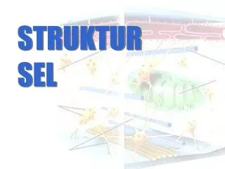
STRUKTUR SEL TUMBUHAN. Perbedaan dengan sel hewan:. 1. Adanya dinding sel yang tebal. 2. Adanya plasmodesmata. 3. Vakuola besar. 4. Badan mikro. 5. Diktiosom. 6. Kloroplas. KOMPONEN PROTOPLASMA. Struktur sel tumbuhan terdiri atas:. 1. Dinding Sel. Bahan polisakarida (selulosa).

E N D
STRUKTUR SEL TUMBUHAN Perbedaan dengan sel hewan: 1. Adanya dinding sel yang tebal 2. Adanya plasmodesmata 3. Vakuola besar 4. Badan mikro 5. Diktiosom 6. Kloroplas
Struktur sel tumbuhan terdiri atas: 1. Dinding Sel Bahan polisakarida (selulosa) Fungsi: melindungi membran sitoplasma dan sitoplasma
Karakter Dinding Sel Tumbuhan - Fleksible dan terkadang sangat padat - Posisi di luar membran sel - Sebagai pendukung, pelindung, penyaring, dan sebagai penekan pembuluh untuk mencegah over expansion saat air masuk sel - Fleksible dan terkadang sangat padat - Dinding primer bersifat semipermeabel,
3 Wedge-shaped expansins, activated by low pH, separate cellulose microfibrils from cross-linking polysaccharides. The exposed cross-linking polysaccharides are now more accessible to cell wall enzymes. 4 The enzymatic cleavingof the cross-linking polysaccharides allowsthe microfibrils to slide.The extensibility of thecell wall is increased. Turgorcauses the cell to expand. 2 The cell wallbecomes moreacidic. 1 Auxinincreases theactivity ofproton pumps. 5 With the cellulose loosened, the cell can elongate. Cell wallenzymes • Cell elongation in response to auxin Expansin Cross-linkingcell wallpolysaccharides CELL WALL Microfibril H2O Cell wall Plasma membrane H+ H+ H+ H+ H+ H+ H+ H+ Cytoplasm Nucleus Vacuole ATP Plasma membrane H+ Cytoplasm
Dinding Sel Primer - mengalami pemanjangan - mengandung sedikit lignin dan ikatan silang fenol • Beberapa fungsi dinding sel primer: • Pendukung struktural dan mekanik • Mengatur dan menetukan ukuran sel • Menahan tekanan internal turgor sel • Mengendalikan laju dan arah pertumbuhan • Bertanggung jawab terhadap arsitektur dan bentuk tumbuhan • Mengatur difusi bahan melalui apoplas • Penyimpan karbohidrat – dinding biji dapat dimetabolisme • Pelindung dari patogen, dehidrasi, and faktor lingkungan • Sumber aktif molekul sinyal biologi • Interaksi antar sel
Key Symplast Apoplast Transmembrane route Apoplast The symplast is the continuum of cytosol connected by plasmodesmata. The apoplast is the continuum of cell walls and extracellular spaces. Symplast Symplastic route Apoplastic route Transport routes between cells.At the tissue level, there are three passages: the transmembrane, symplastic, and apoplastic routes. Substances may transfer from one route to another.
Dinding Sel Sekunder - Tidak mengalami pemanjangan - Mengandung banyak lignin - Tumbuh ke arah dalam, persis di atas membran sel
Beberapa reaksi mikrokimia terhadap dinding sel : 1) Selulosa S + ZnCl-J → ungu S + JKJ + H2SO4 → biru 2) Hemiselulosa HS + ZnCl-J → biru pucat 3) Lignin Zat kayu yang terdapat pada dinding sel yang telah mengkayu. L + ZnCl-J → kuning L + anilin + H2SO4 → kuning L + floroglusin + asam pikrat → merah L + fuchsin + asam pikrat → merah 4) Suberin Terdapat pada dinding sel gabus S + sudan III → merah S + ZnCl-J → coklat S + KOH → kuning
5) Protopektin P + ZnCl-J → kuning coklat P + asam encer larut dalam alkali 6) Pektin Dapat ditemukan pada dinding sel dari buah yang mengandung banyak gula. Bila buah dimasak tampak beberapa zat gelatin 7) Kitin Dapat ditemukan pada dinding sel Fungi (jamur) 8) Kersik (SiO2) Pada dinding sel batang Gramineae, Cyperaceae, Equisetinae, Diatomae 9) Kapur Misal pada dinding sel ganggang Chara sp
PENEBALAN DINDING SEL Menurut cara penebalannya, dapat terjadi secara : APOSISI Yaitu dengan cara menempelkan/melapis-lapiskan bahan penebalan (zat selulosa) pada lamela tengah (substansi interseluler), biasanya pada dinding primer. Contoh : sel parenkim, floem INTUSUSEPSI Penebalan yang terjadi dengan menyisipkan bahan-bahan penebalan di antara mikrofibril
Menurut arah penebalannya : SENTRIPETAL : Yaitu penebalan ke arah pusat sel/dalam. Contoh : pada sel epidermis daun beringin (Ficus sp), terdapat tangkai selulosa yang akan memanjang dan kemudian dideposisikan zat CaCO3 yang makin lama makin banyak sel akan melebar dan disebut litokis. Penebalannya disebut sistolit. SENTRIFUGAL Yaitu penebalan ke arah luar. Contoh : ─ pada polen (ss), terdapat tonjolan-tonjolan yang merupakan penebalan ke arah luar. ─ pada rambut daun (trikoma), misal : daun Artocarpus communis mempunyai rambut-rambut pelindung pada daunnya. Penebalannya terjadi secara intususepsi.
According to the accepted current theory, known as the fluid mosaic model, the plasma membrane is composed of a double layer (bilayer) of lipids, oily substances found in all cells (see Figure 1). Most of the lipids in the bilayer can be more precisely described as phospholipids, that is, lipids that feature a phosphate group at one end of each molecule. Phospholipids are characteristically hydrophilic ("water-loving") at their phosphate ends and hydrophobic ("water-fearing") along their lipid tail regions. In each layer of a plasma membrane, the hydrophobic lipid tails are oriented inwards and the hydrophilic phosphate groups are aligned so they face outwards, either toward the aqueous cytosol of the cell or the outside environment. Phospholipids tend to spontaneously aggregate by this mechanism whenever they are exposed to water.
2. Plastida Diameter 4 – 6 mikron Berwarna (kromoplas) dan tidak berwarna (leukoplas) Leukoplas: amiloplas & lipoplas Kromoplas yang mengandung klorofil adalah kloroplas
Plastida dikenal dalam berbagai bentuk: • proplastida, bentuk belum "dewasa" • leukoplas, bentuk dewasa tanpa mengandung pigmen, ditemukan terutama di akar • kloroplas, bentuk aktif yang mengandung pigmenklorofil, ditemukan pada daun, bunga, dan bagian-bagian berwarna hijau lainnya • kromoplas, bentuk aktif yang mengandung pigmen karotena, ditemukan terutama pada bunga dan bagian lain berwarna jingga • amiloplas, bentuk semi-aktif yang mengandung butir-butir tepung, ditemukan pada bagian tumbuhan yang menyimpan cadangan energi dalam bentuk tepung, seperti akar, rimpang, dan batang (umbi) serta biji. • elaioplas, bentuk semi-aktif yang mengandung tetes-tetes minyak/lemak pada beberapa jaringan penyimpan minyak, seperti endospermium (pada biji) • etioplas, bentuk semi-aktif yang merupakan bentuk adaptasi kloroplas terhadap lingkungan kurang cahaya; etioplas dapat segera aktif dengan membentuk klorofil hanya dalam beberapa jam, begitu mendapat cukup pencahayaan.
3. Vakuola Pada sel tumbuhan besar dan jelas Vakuola tumbuhan dikelilingi membran tunggal (tonoplas) Berisi air, fenol, antosianin, alkaloid dan protein
druse rafida oksalat silikat Batu ginjal
Angiosperm structure • Three basic organs: • Roots (root system) • fibrous: mat of thin roots • taproot: one large, vertical root • Stems (shoot system) • nodes: leave attachment • internodes: stem segments • axillary bud: dormant, vegetative potential • terminal bud: apex of young shoot • apical dominance: inhibits axillary buds • Leaves (shoot system) • blade • petiole
Summary of primary & secondary growth in a woody a stem PRIMARY PRIMARY LATERAL SECONDARY MERISTEMS TISSUES MERISTEM TISSUES Protoderm Epidermis Secondary phloem Primary phloem Vascular Procambium cambium Secondary Primary xylem xylem Ground meristem Ground Pith & tissue: Cortex Cork cambium Cork Apical meristem of stem Periderm