Download
1 / 22
220 likes | 348 Views
Pre-operative Management of the Complex Neonate with CHD Is it all about BLOOD flow?. CVICU LECTURE SERIES Levine Children’s Hospital Charlotte, North Carolina. Pediatric Congenital Heart Surgery. Heart begins to form at the 5 th week of gestation

E N D
Pre-operative Management of the Complex Neonate with CHDIs it all about BLOOD flow? CVICU LECTURE SERIES Levine Children’s Hospital Charlotte, North Carolina
Pediatric Congenital Heart Surgery • Heart begins to form at the 5th week of gestation • Intracardiac defects occur at time of heart forming inutero • No identified etiology • Infants present at birth if congenital lesion is especially ductal dependant • Surgery on neonates/children preformed • Emergently • Day of life 1-10 • One month of life • > 1mo of age
Congenital Heart Disease • Acyanotic Defects • Atrial Septal Defect • Truncus Arteriosus • Atrioventricular Canal Defect • Ventricular Septal Defect • Tetralogy of Fallot • Aortic Stenosis • Total Anomalous Pulmonary Venous Return • Cyanotic Defects • Pulmonary Atresia/ Tricuspid Atresia • Tetralogy of Fallot with severe pulmonary stenosis • Hypoplastic Left Heart Syndrome • Transposition of the Great Arteries • Obstructed Total Anomalous Venous Return
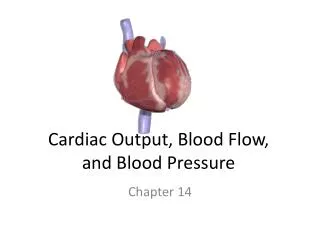
Adjustment after birth • The first breath: … the pulmonary alveoli open up:… pressure in the pulmonary tissues decreases… Blood from the right heart rushes to fill the alveolar capillaries… Pressure in the right side of the heart decreases • … Pressure in the left side of the heart increases as more blood is returned from the well-vascularized pulmonary tissue via the pulmonary veins to the left atrium
Physiology • Pulmonary vascular resistance decreases more than ten fold, thus increasing PBF • Increase of PBF raises left atrial pressure, thus closing the patent foramen ovale • SVR increases • With oxygen induced vasodilatation and lung expansion, PVR decreases to ½ systemic. • After few hours- PDA closes
Differential Diagnosis • Confirm central cyanosis with arterial blood gas (ABG) in room air, if possible, a sample from the right arm is the best site • Correct metabolic acidosis and systemic hypoperfusion if present with fluid boluses and bicarbonate • Check Blood pressure- 4 ex BP • Exam
Hyperoxia Test • 100% FiO2 into headbox for > 10 min • monitor SaO2 • Repeat ABG • PaO2 > 100mmHg or SaO2 increase by 15%: pulmonary disease likely • PaO2 < 70mmHg, rise by < 30mmHg or SaO2 unchanged: cardiac cause or PPHN likely • Total Anomalous Pulmonary Venous Drainage (TAPVD) and Hypoplastic Left Heart syndrome may respond • Pulmonary disease with a massive intrapulmonary shunt may not respond
Diagnostic Data • Precise anatomic diagnosis of the patient’s cardiac defect(s). • Pathophysiological effects of the defect on the CV system and other organs prior to surgery. • Patient’s noncardiac medical and surgical history plus the preoperative medication regimen.
Prostaglandin E1 • Significant decrease in morbidity and mortality since 1970’s • Produces vasodilation • Inhibits platelet aggregation • Stimulates intestinal and uterine smooth muscle. • Dose- 0.01mkm- 1mkm • Side effects- Apnea, Hypotension, Hyperthermia, skin flushing
Confirm Diagnosis • Echocardiogram obtained • Confirmation of anatomy- 2 cardiologists • Plan of care • Medical Management • Timing of surgery
Qp:Qs • Pulmonary blood flow vs. Systemic Blood Flow • Normal is 1: 1 • Need to maximize pulmonary blood flow vs. systemic and how to make it “normal”.
Truncus Arteriosus Defect Absence of part or all of the aorticopulmonaryseptum. There could be a partial truncus or a complete absence of the septum that the PA arise from the common trunk. There is an accompanying interventricular defect which results from the lack of a contribution of the bulbar septum to the IV septum. http://www.heartpoint.com/congtruncus.html
Truncus Arteriosus • May be quite stable • Ok to feed if not concerned with increased pulmonary blood flow and decreased systemic blood flow • Rarely need oxygen, could be contraindicated. • Usually ready for surgery at 2 weeks of age, once PVR drops.
Transposition of the Great Arteries Improper spiraling of the Aorticopulmonary septum.
Transposition of the Great Arteries • Need Balloon Atrial Septostomy if no ASD and patient is cyanotic, or Small VSD with “streaming effect”. • Can be done at bedside/ cath lab. • Pt kept on PGE until BAS performed • After BAS can DC PGE if patient stable, yet would go slow! • Await 1-3 days post BAS for surgery • Ok to feed and wait if doing well
Hypoplastic Left Ventricle Improper development of the semilunar AV valves, and improper remodeling of the AV endocardial cushion tissues. This results in the mitral valve becoming stenotic or atretic as well as atretic aortic valve causing hypoplasia of the arch.
Management of HLHS • ABG : PH 7.40,pa02 40, PC02 40 • If any restriction to the atrial septum, usually baby is well balanced. • If ASD is predominantly with L-R shunting at atrial level – Qp: Qs 2: 1 • Need to decrease pulmonary blood flow • Hypoxic air mixture • Hypoventilation
Management of HLHS • Prostaglandin –start 0.03mkm • Dopamine/Milrinone if necessary to keep MAP 40-50. • NPO- center dependant • If going to feed- use trophic feeds • Watch BUN/CRE closely • Lactic Acid- if slowly increasing- may need to go to surgery for palliation sooner • NIRS- assess preop • Usual time for surgery 3-7 days of age.
Tetralogy of Fallot Unequal partitioning of the aorta and PA leaves the PA narrowed. Causing 4 characteristics -pulmonary stenosis -a failure of the aorta to shift -interventricular septal defect -RV hypertrophy
Tetralogy of Fallot (severe PS) • Patient usually kept on PGE due to cyanosis • Pulmonary stenosis significant. • Cardiac Cath- to balloon PV if able • If unsuccessful- continue PGE • Stable on room air, or O2 • Placement of Blalock Taussig Shunt for PBF and ligation of PDA. • Neonatal Repair?
Icu Management • Critically depends on cardiac lesion • Diagnosis is crucial • Utilize Diagnostic Imaging • Trend Lactic’s, Pa02, physical exam • Communication is key with Cardiology and Intensive Care
Physiology • Understanding Qp: Qs can allow management strategy to affect outcomes. • Utilize PGE whenever unsure • Ask , ask and ask again if differential does not add up!