Download
1 / 34
350 likes | 382 Views
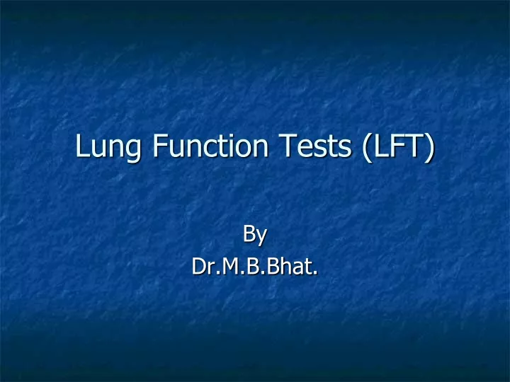
Lung Function Tests (LFT). By Dr.M.B.Bhat. Pulmonary Volumes & Capacities. Volumes –Simple volume obtained only by recording from the instrument Capacities – Consists of more than one volume; Obtained either directly by recording or by addition of different volumes

E N D
Lung Function Tests (LFT) By Dr.M.B.Bhat.
Pulmonary Volumes & Capacities • Volumes –Simple volume obtained only by recording from the instrument • Capacities – Consists of more than one volume; Obtained either directly by recording or by addition of different volumes • Method of study by – Spirometry • Apparatus used – Spirometer (Spirograph) • Record obtained -- Spirogram
Lung Volumes • Tidal volume (TV) –500ml • Inspiratory reserve volume (IRV) –3100ml • Expiratory reserve volume (ERV) –1200ml • Residual volume (RV) –1200ml • Significance of RV – • Prevent collapse of alveoli • Ensuring continuous Blood-Gas exchange • Buffering of alveoli gas – Keep the composition of alveolar gases constant
Pulmonary Capacities • Inspiratory capacity (IC) –IRV + TV =3600ml • Functional residual capacity (FRC) or Functional residual volume (FRV) –RV + ERV = 2400ml • Vital capacity (VC) or Forced Vital capacity (FVC) –IRV + TV (IC) + ERV = 4800ml • Total lung capacity (TLC) -- IRV + TV (IC) + ERV + RV = 6000ml (6 liters)
Variations • All lung volumes & Capacities can – • Vary depending upon sex, size & body build of the individual • It increases proportionate to size • About 20 to 25% less in female than male • More in athletic & large persons
Limitation of spirometry • With spirometry – the volumes & capacities that can be expired out only canbe measured • Hence, the volume which cannot be measured by spirometry are– • Residual volume • So, also, FRC & TLC
Measurement of RV • Open circuit method (Nitrogen washed out method) • Closed circuit method (Helium method) • By Body Plethysmograph method
N2 Washed out method • Start experiment after forceful expiration – • By inspiring pure O2 & expire the gas into Douglas bag –for 10 minutes • (In normal person, all N2 present in the alveoli is washed out within 2 minutes. Even in disease person (asthma Or emphysema) all N2 present in the alveoli is washed out within 7 minutes)
Calculation • Volume of air collected in Douglas bag at the end of experiment– (say 60 liters) • Concentration of N2 in collected air –(2%) • Total volume of N2 in collected air = Total volume of air X conc. Of N2 (60 X 2%) • Residual volume = Total volume of N2/ concentration of N2 in atmospheric air (80%) • = (60 X 2%)100/80
Helium method • Keep the known concentration of Helium gas mixture (of known volume) in the chamber & during experiment period, the subject rebreath in & out with the chamber (for 10 minutes) • Calculation –RV = {(Ci He/C2 He) –1} V1 • Ci He = Initial conc. Of He in the chamber • C2 He = Final conc. Of He in the chamber • V1 = Volume of gases in the chamber
Body Plethysmograph • Based on Boyle’s law -- PV = P1V1 • P = Alveolar pressure; • V= Volume of gas in alveoli –to be found out • First measure P ; start experiment by inspiring known volume (∆V) gas & measure new P (P1) • Calculation = • PV = P1 (V + ∆V) ; PV = P1 V + P1 ∆V • PV –P1 V = P1 ∆V ; V (P –P1) = P1 ∆V • V = P1 ∆V / (P –P1)
All three methods, depending upon the starting condition of the experiment, the following volume can be obtained – After normal expiration – FRC After forceful expiration –RV After maximal inspiration – TLC Thoracic gas volume – the volume of gas in thorax, including gas in the non-communicable alveoli. In normal person it is equal to FRC It canbe measured only by body plethysmograph
Dead Space Volume (DSV) • Definition • Function – Purification, Humidification & Air-conditioning • Types –2 • Anatomical dead space volume– volume of gas in respiratory tract ( up to terminal bronchiole). Normal value – 150ml • Physiological dead space volume (Total dead space volume) – • Anatomical DSV + Volume of gas in unperfused alveoli in Normal person – same as Anatomical DSV • Normal healthy person – • Physiological DSV = Anatomical DSV
Determination of Anatomical DSV(Fowler’s method –Single breathe pure O2 –Nitrogen measurement)
Physiological dead space volume • Measurement by using – • Bohr’s formula (Bohr’s equation)–
Dynamic Lung volume & Capacities (or) Lung Function tests • 1. Respiratory minute volume (RMV) or Pulmonary ventilation = TV X RR • (Range of TV –Up to ½ of VC; Normal range of RR –12 to 18; varies from 4 to 40/min) • 2. Alveolar ventilation = (TV – DSV) X RR
3. Maximum Voluntary Ventilation (MVV) orMaximum Breathing capacity (MBC) = 80 to 120L/min Determination –With maximum voluntary effort breath as deep & as quick as possible for 15 sec (reason?), the volume obtained is multiplied by 4 & expressdper minute. Varies with age, sex & body size Also depend on – muscular force available, compliance of thoracic wall & lungs, air way resistance.
4. Pulmonary reserve or Breathing reserve = MVV – RMV 5. dyspnoeicIndex (DI) – Pulmonary reserve expressed as % of MVV (MVV – RMV)/MVV X 100 When DI is < 60 to 70% dyspnoea present
6. PEFR (Peak expiratory flow rate)-- >400L/min (respiratory Muscle endurance) 7. MMEFR (Maximal mid-expiratory flow rate -- > 300L/min (sensitive indicator of small air way diseases; so in obstructive diseases it is decreased) (Both are measured by Wright’s flow meters)
8. Timed Vital Capacity (TVC) or Forced Vital Capacity (FVC) –VC measured at each unit time (second). Accordingly, volume expired each unit and expressed as % of TVC; as -- FEV1 –80 to 85% FEV2– 90 to 95% FEV3 -- >97% Helps to differentiate Restrictive with Obstructive type of respiratory diseases. In both types of diseases -- Flow rates & MVV Restrictive disorders -- VC; FEV1 normal (Pulmonary fibrosis; Kyphoscoliosis; Ankyosis spondylitis) Obstructive diseases –VC normal & FEV1 (Bronchial asthma; Emphysema)
Some Respiratory terms • Eupnoea – Normal Breathing • Dyspnea – difficult or labored breathing in which the subject is conscious of shortness of breathing • Hyperpnoea – increase in rate & depth of breathing regardless of subject’s subjective sensation • Normal individual is not conscious of respiration until ventilation is doubled. • Ventilation is not uncomfortable until it is > tripled or quadrupled • Tachypnea –is rapid shallow breathing • Hypercapnia -- CO2….. • Hypoxia –O2
Alveolar air sampling • By Haldane & Priestly method • Tube of 120cm long & 2.5cm wide with evacuated gas sampling side tube • Last 10ml of expired air collected as sample of alveolar gas
Gas analysis • Lloyd's modification of Haldane’s volumetric apparatus --procedure Gas sample (known volume) – pass through KOH solution (CO2 absorbed) –pass through alkaline pyrogallol solution (O2 absorbed) Oxymeter –Para-magnetic properties of O2 CO & CO2 analyzer –Infra-red absorption (spectroscopic method) Nitrogen meter –N2 emits light in electric field in vacuum Caltharmometry –using thermal properties of gases Gas-chromatography & mass spectrometry
Blood-Gas analysis • Van Slyke’s apparatus – • Blood + acidified, air-free ferric cyanide solution is subjected to vacuum. • Acid –drive off CO2 • Ferric cyanide – drive off O2 • Vacuum –drive off N2 • Haldane Blood-gas apparatus – • Oxalated blood is treated with sapoin (for hemolysis), then Potassium ferric cyanide is added to liberated O2.